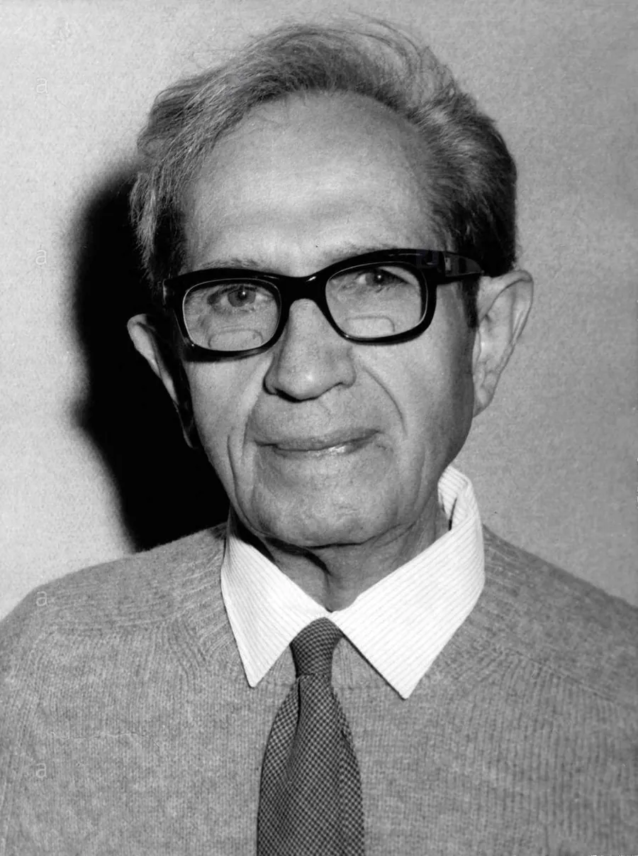
George Emil Palade, 1912–2008
A Romanian doctor timed a protein's journey through the cell and won a Nobel Prize
In 1953 George Palade showed that almost every kind of cell he examined holds tiny grains, many of them stuck to membranes: the ribosomes, where proteins are made. Then, with Lucien Caro and James Jamieson, he measured how many minutes new proteins take from the ribosome to the granules from which they leave the cell. The name ribosome was proposed at a meeting in 1958, and others had seen similar particles before him.
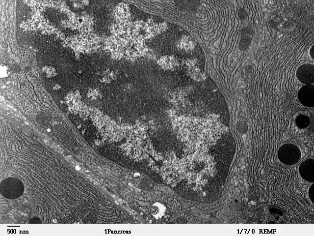
If a cell were as tall as you
A cell from a rat's pancreas, of the kind Palade studied in guinea pigs, is about 15 micrometres across, counted as a sphere of the same volume. Enter your height and see how big its parts would be at the same magnification.
- a ribosome would be
- 2.8 mm
- the 1955 grains, as they looked on sections
- 1.0–1.5 mm
- a granule of enzymes
- 7 cm
- 15-micrometre cells end to end, to your height
- 101,903 cells
Schematic drawing, after the structures Palade names in his Nobel lecture. The ribosomes are enlarged so that they can be seen.
1 Iași, 19 November 1912
A philosophy professor's son chooses medicine
1912
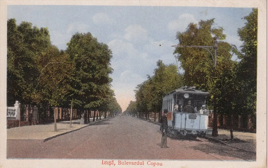
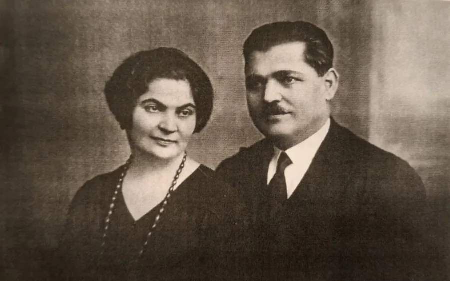
George Emil Palade was born in Iași, in Romania, on 19 November 1912. His father, Emil Palade, was a professor of philosophy; his mother, Constanța Cantemir-Palade, was a teacher. In the autobiography he wrote for the Nobel Foundation, Palade says that from them he learned early a great respect for books, scholars and education.
He started school in Iași and took his baccalaureate at the Hasdeu lyceum in Buzău. His father hoped he would study philosophy, as he had. The son, Palade writes, “preferred to deal with tangibles and specifics”, and in 1930 he entered the School of Medicine in Bucharest.
From his first years there he was drawn to the basic sciences behind medicine, after talking with his professors, Francisc Rainer in anatomy and André Boivin in biochemistry. As a student he was already working in the anatomy laboratory. He still did six years of hospital training, mostly in internal medicine. For his doctorate, though, he chose a subject unusual for a future doctor.
After graduating in 1940 he spent a short time as an assistant in internal medicine, then went back to anatomy. His reason: the gap between what doctors of the time knew and what was expected of them made him “rather uneasy”.


Choose, then see what the sources say
What was Palade's medical doctorate about?
The dolphin's kidney. The thesis, defended in 1940, was called “The urinary tubule of the dolphin” and tried to explain how a mammal's kidney had adapted to life in the sea. Marilyn Farquhar, a cell biologist and his second wife, writes that the kidneys came from the Black Sea and that Palade's three-dimensional model stayed on display in the faculty's Anatomical Museum, where for many years it inspired generations of students.
2 Bucharest – New York, after the war
One journey, dates that do not agree
1946
In 1943 Palade had won, by competition, the post of associate professor of anatomy in Bucharest. During the war he served as a doctor in the Romanian army. After the war his professor, Grigore T. Popa, urged him to go and study in the United States, and Farquhar writes that Popa gave him several letters of recommendation to American laboratories.
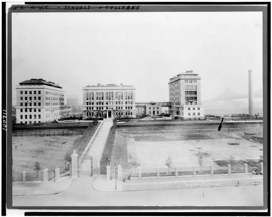
He spent his first months in America at New York University, as a visiting investigator in Robert Chambers's biology laboratory. There he heard Albert Claude, a Belgian at the Rockefeller Institute, give a seminar on what he could see with the electron microscope. Palade writes that he was “fascinated” and “extremely happy” when, after a short discussion, Claude asked him to come and work with him from the autumn of the same year. Chambers was retiring that summer.
At the Rockefeller, in the mid-1940s, the two methods Palade would use for the rest of his life were being worked out: looking at cells with the electron microscope, and taking a cell apart by spinning it in a centrifuge.

Choose, then see what the sources say
In which year did Palade arrive in America?
It depends on whom you read. Palade himself writes, in his 1974 autobiography, that he came to the United States “in 1946 for further studies”. James Jamieson, a former colleague, writes 1945. Marilyn Farquhar says, in her memoir for the US National Academy of Sciences, that he bought a passport and a visa on the black market, slipped out of the country at night and sailed for New York from Casablanca at the end of 1945, which would fit an arrival in 1946. The Nobel press release says he joined the Rockefeller in 1947, although Palade writes that he went there in the autumn of his first year. None of these sources cites a travel document.
3 New York, 1946–1953
Two new ways to look at a cell
1,250times finer, in principle
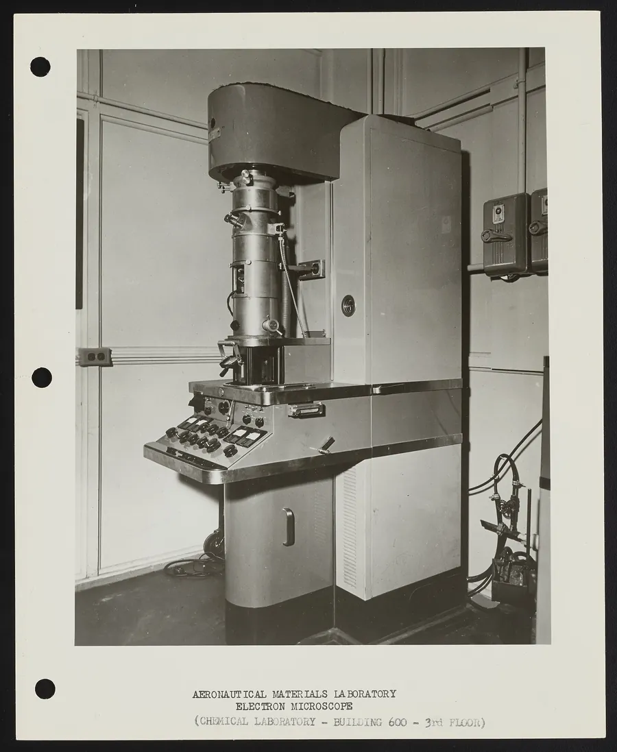
A light microscope cannot distinguish details smaller than about 2,500 ångströms. An ångström is a ten-billionth of a metre, so that is a quarter of a micrometre. The electron microscope uses a beam of electrons instead of light, and Farquhar writes that its practical limit is about 2 ångströms. The two limits differ by a factor of about 1,250. At first, though, it could look only at cells grown in dishes and spread thin. In 1945 Keith Porter, Albert Claude and Ernest Fullam had published pictures of such cells, from chick embryos, and seen in them a network of membranes: the endoplasmic reticulum.
The other method was Claude's. You crush the cell and separate the pieces in a centrifuge, a machine that spins tubes very fast: the heaviest pieces settle first at the bottom of the tube, then, at ever faster spins, the others in turn. In this way Claude obtained four fractions: three successive sediments and the fluid left on top. Palade worked on this first: with George Hogeboom and Walter Schneider he developed the “sucrose method”, which keeps whole the mitochondria, the structures the cell draws its energy from.
In 1949 Claude went back to Belgium. When the head of the department, James Murphy, also retired, Palade and Keith Porter were left “orphans”, as Palade puts it, and were “adopted” by the institute's director, Herbert Gasser. They improved the way tissue was cut and fixed, that is, preserved with chemicals before it is looked at. The novelty of Palade's 1952 fixative, Farquhar writes, “consisted simply” in a buffer, a solution that holds acidity steady, used as the solvent for osmium. With slices no thicker than 0.05 micrometres and an RCA microscope, the cells of any tissue could at last be looked into.
What comes out of the centrifuge
Pick a fraction to see what it holds and where it sits in the cell.
Nuclei. The heaviest pieces, first to the bottom of the tube: the nuclei, which hold the cell's genetic material.
Mitochondria. The mitochondria, which the Nobel press release calls the cell's energy producers.
Microsomes. The microsomes: small pieces of membrane. Palade and Siekevitz will show in 1956 that they are fragments of endoplasmic reticulum.
Cell sap. What is left on top: the cell's fluid, with everything that has not settled.
De Duve later found enzymes that fitted none of the four. They sat in a fifth fraction, the lysosomes.


Choose, then see what the sources say
On the membranes of the reticulum Palade will find tiny grains. In which of Claude's four fractions would they end up, membranes and all?
In the microsomes. In 1956 Palade and Philip Siekevitz showed that the microsomes of the pancreas are pieces of endoplasmic reticulum, with the grains still attached to the outside of the membrane. Grains that have come loose can then be collected with one more spin.
4 Pocono Manor, 1953 · New York, 1955
The grains on the membranes
150ångströms, the top of the usual range
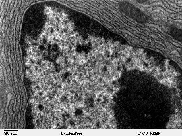
In pictures taken at magnifications of 5,000 to 10,000 times and then enlarged again on paper, Palade saw something that recurred in almost every kind of cell he examined: small, dense, round grains, most of them 100 to 150 ångströms across, or 10 to 15 nanometres. A nanometre is a millionth of a millimetre.
In specialised cells, such as those of the pancreas, the grains sat on the membranes of the endoplasmic reticulum, usually in no particular order, sometimes in short rows. In cells that divide quickly they floated free. Palade presented his observations in 1953 at the annual meeting of the Electron Microscope Society of America, at Pocono Manor in Pennsylvania. The paper reached the journal on 15 October 1954 and came out on 25 January 1955, in the first issue of a new journal. The two dates used today, 1953 by the Romanian Academy and 1955 by the Nobel Foundation, match the talk and the paper.
The paper is careful. Palade first asks whether the grains might be an artefact, something made by the fixing, and shows that he sees them with another fixative too. He notes that they appear where the cytoplasm, everything in the cell outside the nucleus, stains strongly because of ribonucleic acid, RNA, a molecule related to DNA. But he writes that grouping them under one name rests “exclusively on morphological findings”, meaning on how they look, and that the “final proof” of the link with RNA “can be expected in the future”.
In the same paper he cites those who had seen something similar: Keith Porter in 1952, “macromolecular units”; Sjöstrand and Rhodin in 1953, “dense dots”; Slautterback, “small microsomes”. And biochemists, Petermann and her colleagues, Barnum and Huseby, had already isolated RNA-rich particles of about the same size by centrifuging. What Palade showed was that the grains appear in all of the more than 40 kinds of cell he examined, in mammals and birds, with a single exception, the adult red blood cell, and that in many cells they sit on the membranes of the reticulum.

Choose, then see what the sources say
What does Palade's 1955 paper say these grains do?
It does not say. He links them with RNA and leaves the proof for later, and the paper says nothing about making proteins: the word appears only inside “nucleoprotein”, about the particles Petermann isolated. The Nobel Foundation's page now says that in 1955 Palade discovered “previously unknown organelles”, ribosomes, “where the cell's formation of proteins takes place”. His own paper shows that similar particles had been seen and isolated before, and says nothing about their role. His contribution was the systematic description of the particles; their role was proved later.
5 New York · Cambridge, Massachusetts, 1955–1960
A name and a proof
1958
In 1955 the biochemist Philip Siekevitz joined the laboratory. He had built one of the first systems in which proteins are made in a test tube. Together they chose the guinea pig's pancreas and used both methods at once: they separated the cell into fractions by centrifuging and checked under the microscope what each one held. That is how they showed, in 1956, that the microsomes of the pancreas come mainly from the rough reticulum, broken into pieces when the cell is crushed, and that the grains on them are ribonucleoprotein particles, made of RNA and protein.
What the particles did was still to be proved. In 1960 Kirsch, Siekevitz and Palade showed that particles detached from guinea-pig liver microsomes build amino acids into proteins, even in a test tube. The 1974 Nobel press release says Palade showed this “with other groups”.
Before today's name, the particles had been called several things: the “small particulate component of the cytoplasm”, as Palade called them in 1955, then “ribonucleoprotein particles”, and for a short while, Farquhar writes, “Palade granules”.
Choose, then see what the sources say
Where, in the sources cited, does the word “ribosome” first appear?
At the 1958 symposium. In February 1958 the Biophysical Society held its first symposium, at the Massachusetts Institute of Technology, on microsomal particles and protein synthesis. In his introduction to the volume, R. B. Roberts writes that participants did not agree on what “microsomes” meant and that “during the meeting the word ‘ribosome’ was suggested”; it “has a pleasant sound”. He does not say who suggested it, so the sources do not show whether it was Palade or not. In his Nobel lecture, Palade cites Roberts's introduction for the term.
6 New York, 1964–1967
On the trail of a protein
60minutes to the granules
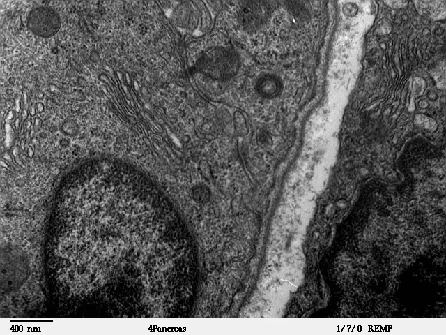
A pancreas cell makes the enzymes of digestion and sends them out into the ducts that lead to the gut. Palade calls it “a very efficient protein producer”. Of its pictures he writes that they had for him “the effect of the song of a mermaid: irresistible and half transparent”.
The centrifuge did not separate the cell's pieces cleanly, so Palade and Lucien Caro tried something else. They injected guinea pigs with radioactive leucine, an amino acid the cell builds proteins from. Proteins made in the next few minutes thus carried a radioactive label. After a few minutes, a few tens of minutes or an hour, they fixed the tissue and looked under the microscope for the marks the radioactivity had left on a photographic layer. The method is called autoradiography.
In 1967 James Jamieson and Palade followed the same route in slices of pancreas kept alive in a dish, mostly by separating the cells into fractions: three minutes with the radioactive amino acid, then a nutrient solution without the label. The new proteins moved from the reticulum into the small vesicles at the edge of the Golgi apparatus, where the labelled protein peaked seven minutes after the solution was changed. In autoradiographs, the granules themselves were first seen labelled in the sample taken 57 minutes after the pulse. The times in slices and in the living animal cannot be compared directly: they are different experiments.
Put the stages in order
Which way does a protein made for export travel? Press the stages in the order you think it passes through them, from first to last.
Your order
The order Palade and his colleagues found
- Ribosomes on the reticulum The protein is made here, on the ribosomes attached to the membrane of the reticulum.
- Inside the reticulum≈ 5 min The growing chain passes through the membrane into the inside of the reticulum.
- Vesicles at the edge of the Golgi apparatus Small bubbles of membrane carry the proteins to the Golgi apparatus.
- Condensing vacuoles≈ 20 min In the Golgi apparatus and the vacuoles on its far side, the proteins are concentrated.
- Zymogen granules≈ 60 min The vacuoles become granules, the stores in which the enzymes wait.
- Out, into the lumen When they are needed, the granules empty into the lumen, the space in the middle of a cluster of cells, from where the enzymes leave for the gut.
The minutes are those of 1964, in the living guinea pig: the label is in the reticulum at about 5 minutes, in the Golgi apparatus at about 20 and in the granules after an hour.

Choose, then see what the sources say
What happens to the proteins if the cell stops making energy?
They stay in the reticulum. Palade calls the result “unexpected”: transport to the Golgi apparatus needs energy, which the cell gets from ATP. When the cell makes ATP again, transport resumes.
7 Stockholm, October–December 1974
One prize in three parts
3laureates, a third each
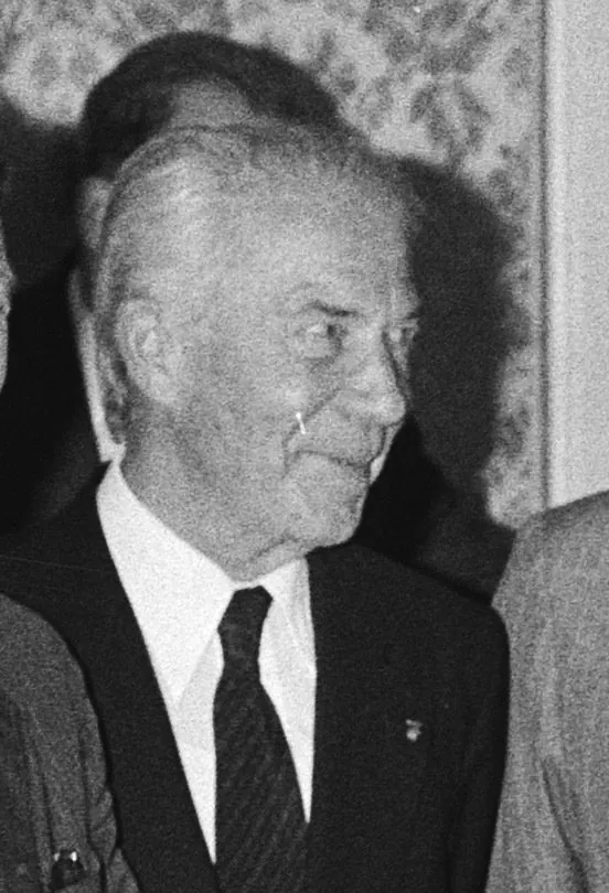
In October 1974 the Karolinska Institutet announced the Nobel Prize in Physiology or Medicine, split into three equal parts: Albert Claude, Christian de Duve and George E. Palade, “for their discoveries concerning the structural and functional organization of the cell”. Palade had been a professor at Yale for a year.
The press release divides the credit. Claude brought the electron microscope to the study of animal cells and developed fractionation by centrifuge. De Duve, starting from Claude's fractions, found enzymes that fitted none of them and arrived at a fifth: the lysosomes, the small sacs in which the cell breaks down its waste. Palade showed how proteins for export pass from the reticulum into the Golgi apparatus, where they are made ready to leave.
At the ceremony Jan-Erik Edström said Palade had developed electron microscopy “to the highest degree of artistry”. In his Nobel lecture, on 12 December, Palade broke secretion into six steps: synthesis, segregation, transport, concentration, storage and discharge. For each he weighed the evidence and said where it was still weak.

Choose, then see what the sources say
Of all the Nobel laureates born on the territory of today's Romania, where does Palade come by year of award?
First. The Nobel Foundation's database lists four laureates born on the territory of today's Romania: Palade in 1974, Elie Wiesel (peace) in 1986, Herta Müller (literature) in 2009 and Stefan Hell (chemistry) in 2014. Palade received the prize as a professor at Yale, and the foundation lists him under the American university.
8 Bucharest · New Haven · La Jolla, 1975–2008
Home and away
1979
On 31 March 1975 the Romanian Academy elected him an honorary member. With Nicolae and Maia Simionescu, two Romanian scientists who worked with him at the Rockefeller and then at Yale, he studied the walls of the smallest blood vessels, the capillaries. Farquhar writes that the Institute of Cell Biology and Pathology in Bucharest was “the dream of the Simionescus”, but that Palade's standing made its creation possible, in the Ceaușescu years. In September 1979 many of those who had worked with Palade and the Simionescus at the Rockefeller travelled to Bucharest for its opening symposium.
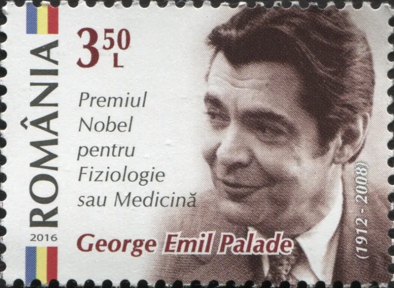
In 1990 he moved to the University of California, San Diego. Farquhar writes that his door was open to everyone, down to the Romanian students who came to meet “their national idol”. In 1994 the Al. I. Cuza University of Iași awarded him an honorary doctorate, and in 2008 he received the Order of the Star of Romania in the rank of Collar. He died on 7 October 2008 at Del Mar, California, aged 95. The Romanian Academy's page gives the date as 8 October.

Choose, then see what the sources say
Which structure in the cell bears Palade's name today?
The Weibel–Palade bodies, rod-shaped granules in the cells that line blood vessels, described by Weibel and Palade in 1964. Farquhar writes that they are the only organelle in the cell that bears his name, although he discovered others. “Palade granules” was a name used briefly for ribosomes.
9 2026
The same cell, today
30nanometres, today's ribosome
After Palade, others found the machinery behind the route he had described. Günter Blobel, who joined Palade's laboratory at the end of the 1960s, proposed in 1971, according to the 1999 Nobel press release, that proteins made for export carry a signal of their own that takes them to the membrane of the reticulum and through it, and in 1975 he showed that the signal is a short string of amino acids. He received the Nobel Prize in 1999.
Venkatraman Ramakrishnan, Thomas Steitz and Ada Yonath won the 2009 Nobel Prize in Chemistry for the structure of the ribosome, atom by atom. The models show that much of its work is done by its RNA; in bacteria, RNA makes up close to two-thirds of the ribosome's mass. In 2013 James Rothman, Randy Schekman and Thomas Südhof were honoured for the machinery that steers vesicles, the bubbles of membrane that carry proteins between the cell's compartments. The prize's scientific background names the 1974 laureates among the pioneers: from Palade's work it was known that proteins leave the reticulum in small vesicles.
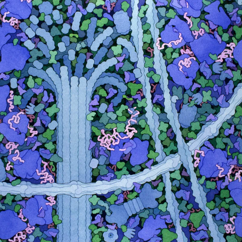
Measured along the three axes of the atomic model of the human ribosome, published in 2015, the ribosome is 26 to 30 nanometres across. The grains in Palade's paper measured 10 to 15. The sizes were measured differently: in 1955, the dense outlines seen on thin, chemically prepared sections; in the atomic model, the full extent of the whole ribosome. The sources cited do not explain the difference.
What the microscope shows
What we know about the same molecules
Who found what
- 1945Porter, Claude, Fullamfirst electron micrographs of whole cells; the endoplasmic reticulum
- 1946Claudeseparating the cell into fractions by centrifuge
- 1952Porter“macromolecular units” in the cytoplasm
- 1953Paladethe grains on the reticulum, presented at Pocono Manor
- 1955Paladethe paper “A small particulate component of the cytoplasm”
- 1956Palade, Siekevitzmicrosomes are pieces of reticulum; the grains are ribonucleoprotein
- 1958the MIT symposiumthe name “ribosome”
- 1960Kirsch, Siekevitz, Paladeparticles detached from microsomes make protein in a test tube
- 1964Caro, Paladethe protein's route, timed in the living guinea pig
- 1967Jamieson, Paladethe route, step by step, in pancreas slices
- 1971Blobelfirst version of the signal hypothesis
- 1974Claude, de Duve, Paladethe Nobel Prize in Physiology or Medicine
- 2009Ramakrishnan, Steitz, Yonaththe structure of the ribosome, Nobel Prize in Chemistry
- 2013Rothman, Schekman, Südhofthe machinery that steers vesicles, Nobel Prize
The story in numbers
| Born | in Iași, on 19 November 1912 |
|---|---|
| Doctoral thesis, 1940 | the dolphin's kidney |
| Leaving for America | Jamieson: 1945; Farquhar: sailed at the end of 1945; Palade: 1946; at the Rockefeller in 1946 or 1947 |
| Electron microscope against light microscope | 1,250 times finer |
| The 1955 grains | 10–15 nanometres, on the membranes of the reticulum |
| The name ribosome | proposed at the MIT symposium, February 1958 |
| A protein's route in the living guinea pig, 1964 | reticulum at about 5 minutes, Golgi at about 20, granules after an hour |
| Without energy | the proteins stay in the reticulum |
| The 1974 Nobel Prize | a third each: Claude, de Duve, Palade |
| The human ribosome, 2015 atomic model | 26–30 nanometres, along its axes |
Quiz: what Palade did and did not do
Eight statements. Choose, then see what the sources say.
The word “ribosome” already appears in Palade's 1955 paper.
False. In 1955 Palade writes of “a small particulate component of the cytoplasm”. The word was proposed at a symposium at MIT in February 1958, and the volume's introduction does not say by whom.
R. B. Roberts (ed.), Microsomal Particles and Protein Synthesis, 1958 (Internet Archive)
In his 1955 paper, Palade writes that the grains make proteins.
False. He links them with RNA, leaves the proof for the future and says nothing about making proteins.
Before him, other researchers had seen or isolated similar particles.
True. His own paper cites Porter, Sjöstrand and Rhodin, Slautterback and the biochemists who had isolated RNA-rich particles.
In the living guinea pig, the radioactive label reaches the Golgi apparatus in about 20 minutes.
True. Caro and Palade find it in the reticulum at about 5 minutes, in the Golgi at about 20 and in the granules after an hour.
L. G. Caro and G. E. Palade, J. Cell Biol. 20 (1964) 473–495
Without energy, the proteins carry on, only more slowly.
False. Without ATP they stay in the reticulum, and transport resumes only when the cell makes energy again.
Palade shared the 1974 Nobel Prize with Albert Claude and Christian de Duve.
True. Each received a third of the prize.
His doctoral thesis was about the dolphin's kidney.
True. “The urinary tubule of the dolphin”, defended in 1940.
Romanian Academy, George Emil Palade, honorary member (in Romanian)
All the sources agree that he arrived in America in 1946.
False. Palade writes 1946, Jamieson 1945, Farquhar says he sailed at the end of 1945, and the Nobel press release puts him at the Rockefeller from 1947.
James D. Jamieson, “A tribute to George E. Palade”, J. Clin. Invest., 2008
What the sources cannot say
- Why the 1955 grains measured 10 to 15 nanometres while today's atomic model of the human ribosome is 26 to 30 along its axes. No source cited explains the gap.
- Who proposed the word “ribosome”. Roberts writes only that it was suggested during the 1958 meeting.
- The exact date of his departure from Romania and the route he took. The only account with a black-market passport and a ship from Casablanca is Marilyn Farquhar's, written after Palade's death, and it cites no document.
- Whether Palade left intending to stay. His autobiography says only that he came “for further studies”.
- Whether the dolphin-kidney model still exists. Farquhar writes that it was on display in the faculty's Anatomical Museum; no source cited says where it is now.
Sources
Palade's life comes from the autobiography he wrote for the Nobel Foundation in 1974, from the memoir Marilyn G. Farquhar wrote for the US National Academy of Sciences and from the Romanian Academy's pages. The science comes from the papers of Palade and his colleagues and from his Nobel lecture. The size of today's ribosome was measured by Marius Comper on the atomic model PDB 4UG0, along its three axes. The size of the cell is the diameter of a sphere with the volume Aughsteen and Cope measured in the rat, 1,670 cubic micrometres.
- George E. Palade, Biographical, Les Prix Nobel 1974 (NobelPrize.org)
- G. E. Palade, Nobel lecture, 12 December 1974 (PDF)
- G. E. Palade, “A small particulate component of the cytoplasm”, J. Biophys. Biochem. Cytol. 1 (1955) 59–68
- G. E. Palade and P. Siekevitz, “Pancreatic microsomes”, J. Biophys. Biochem. Cytol. 2 (1956) 671–690
- R. B. Roberts (ed.), Microsomal Particles and Protein Synthesis, 1958 (Internet Archive)
- L. G. Caro and G. E. Palade, J. Cell Biol. 20 (1964) 473–495
- J. D. Jamieson and G. E. Palade, J. Cell Biol. 34 (1967) 577–596
- J. D. Jamieson and G. E. Palade, J. Cell Biol. 34 (1967) 597–615
- K. R. Porter, A. Claude and E. F. Fullam, J. Exp. Med. 81 (1945) 233–246
- Marilyn G. Farquhar, “George E. Palade 1912–2008”, Biographical Memoirs, National Academy of Sciences, 2017 (PDF)
- James D. Jamieson, “A tribute to George E. Palade”, J. Clin. Invest., 2008
- Karolinska Institutet, press release, Nobel Prize in Physiology or Medicine 1974
- Jan-Erik Edström, award ceremony speech, 1974
- NobelPrize.org, George E. Palade: facts
- Nobel Foundation, laureate database (API 2.1)
- Romanian Academy, George Emil Palade, honorary member (in Romanian)
- Romanian Academy, In memoriam 2008 (in Romanian)
- Karolinska Institutet, Nobel Prize press release 1999 (Günter Blobel)
- Royal Swedish Academy of Sciences, Nobel Prize in Chemistry 2009, press release
- The Nobel Assembly, scientific background to the 2013 prize
- The human ribosome, atomic model PDB 4UG0 (Khatter et al., Nature, 2015)
- A. A. Aughsteen and G. H. Cope, Cell Tissue Res., 1987 (abstract)
- R. Milo and R. Phillips, Cell Biology by the Numbers
The images: public-domain and openly licensed photographs from Wikimedia Commons (author and licence under each image), and a painting by David S. Goodsell, RCSB PDB, under CC BY 4.0. The 1955 micrographs carry no open licence, so the grains are drawn and the plates can be seen in the original paper.