mariuscomper.ukRomână

A world beyond the naked eye

What lives
in a drop
of water?

An alga with dozens of branches is a single cell. A tiny rotifer has organs. Look closer: pond water can hold them both.

Look through the microscope
YES, IT’S A SINGLE CELL.
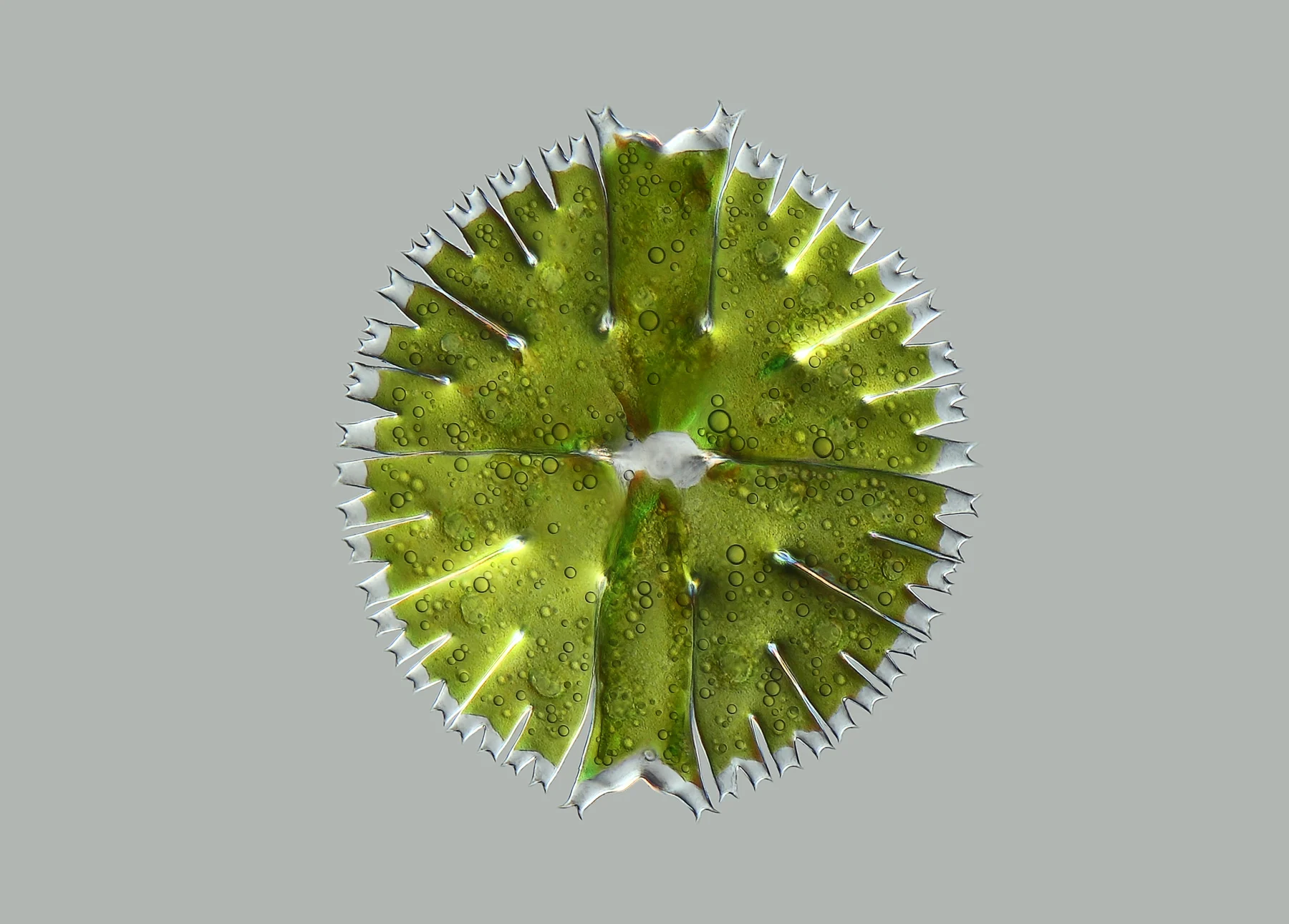
A single green Micrasterias cell with two broad halves and deeply branching edges.
Micrasterias rotata · Anatoly Mikhaltsov · Source · CC BY-SA 4.0

Five life forms, up close

Choose what
to look at.

Change the slide, enlarge the photograph and find the suggested detail. Start with the green alga’s branching edge.

A single green Micrasterias cell with two broad halves and deeply branching edges.
A single-celled green alga photographed under a light microscope. Anatoly Mikhaltsov · Source · CC BY-SA 4.0
View the full photograph ↗

One cell

This entire shape is a single cell.

Micrasterias rotata

Every branch belongs to the same alga. Its two halves meet at a narrow waist. The green comes from chloroplasts, the parts of the cell that capture light for photosynthesis.

LOOK HERE

Follow the edge out to its smallest branches. Then compare the top half with the bottom: they are very similar, without being perfectly identical.
About this organism ↗

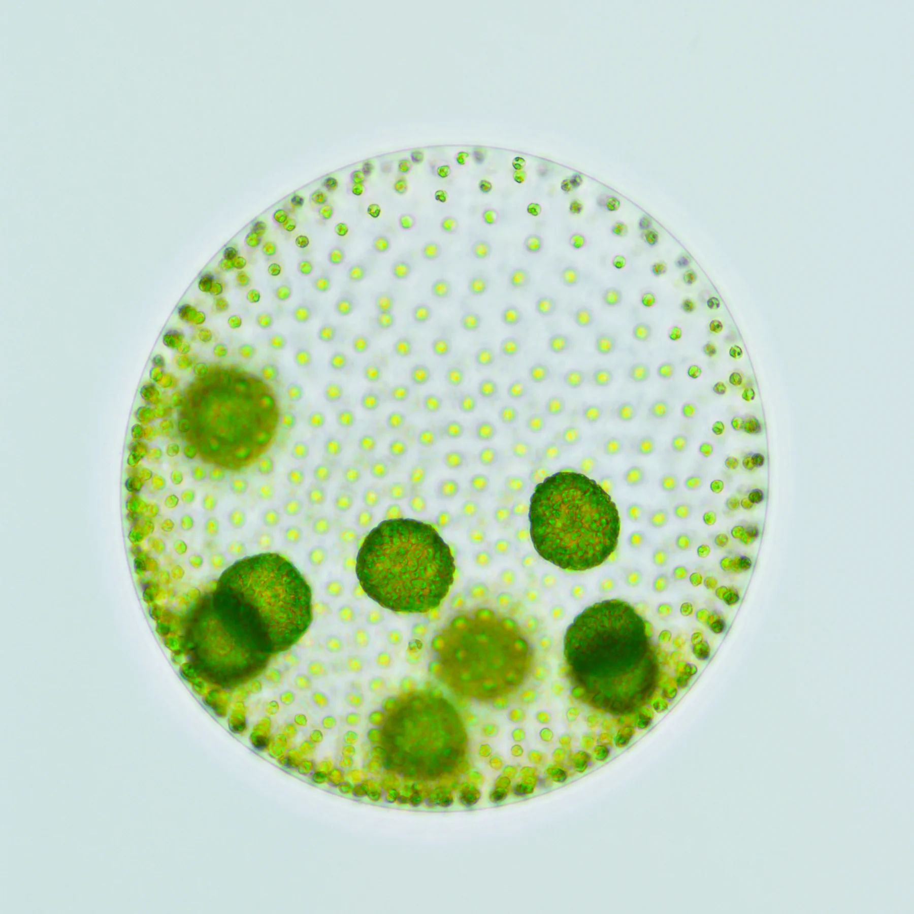
A transparent sphere dotted with tiny green cells, containing several smaller green spheres.
A colony containing daughter colonies. The photograph combines several focus planes. Matthew Herron · Source · CC BY-SA 4.0
View the full photograph ↗

A colony

Inside the big sphere, smaller spheres are growing.

Volvox aureus

The green dots on the surface are cells. Together, they form a Volvox colony. The larger spheres inside are daughter colonies, developing before they are released.

LOOK HERE

Distinguish the tiny surface dots from the green spheres inside. The outer cells have fine filaments called flagella, whose beating propels the colony.

About this organism ↗

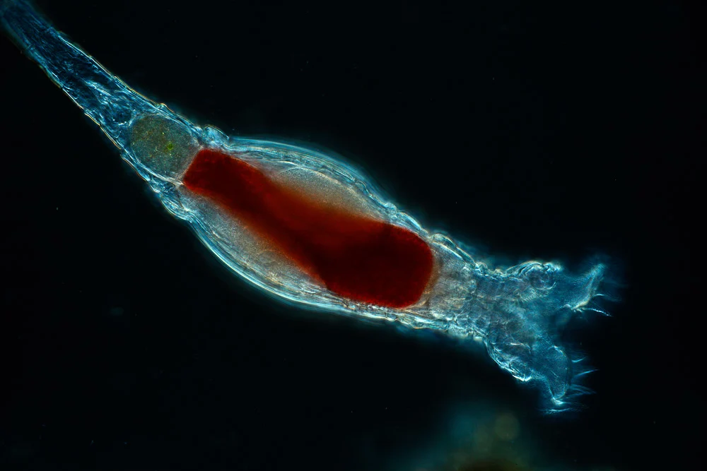
An elongated rotifer against black, with a transparent blue outline, reddish interior and fine cilia on the right.
A rotifer photographed by Frank Fox. The image identification does not specify a species. Frank Fox · www.mikro-foto.de · Source · CC BY-SA 3.0 DE
View the full photograph ↗

An animal

An entire animal, almost transparent.

Rotifera

It has a mouth, a digestive tract and muscles. A rotifer is made of many cells, although it fits in the same microscopic field as single-celled organisms. In many rotifers, cilia around the mouth move water and draw in food particles.

LOOK HERE

At the broad end on the right, look for very fine filaments. Beating one after another, they can look like turning wheels. Further down, you can explore how this illusion works.

About this organism ↗

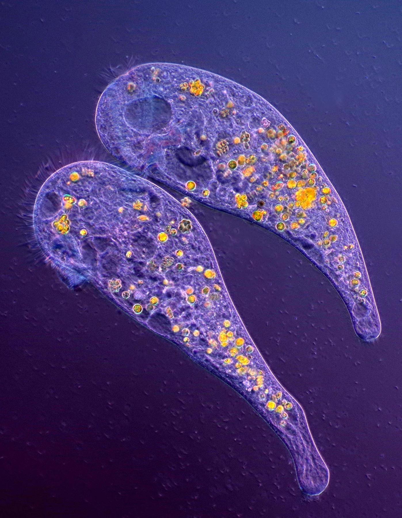
Two violet, trumpet-shaped Stentor cells with small golden inclusions.
Two Stentor cells photographed using polarized light and dark-field illumination. Marek Miś · Source · CC BY 4.0
View the full photograph ↗

One cell

A cell that can rebuild its missing parts.

Stentor

Each of the two shapes in the photograph is one cell. In the species Stentor coeruleus, researchers have observed something remarkable: after a cell is cut in two, each half can regenerate its missing parts if it retains part of the large nucleus.

LOOK HERE

Find the broad end, fringed with cilia. Their beating produces a current that brings in food. The shape can change as the cell contracts.

About this organism ↗

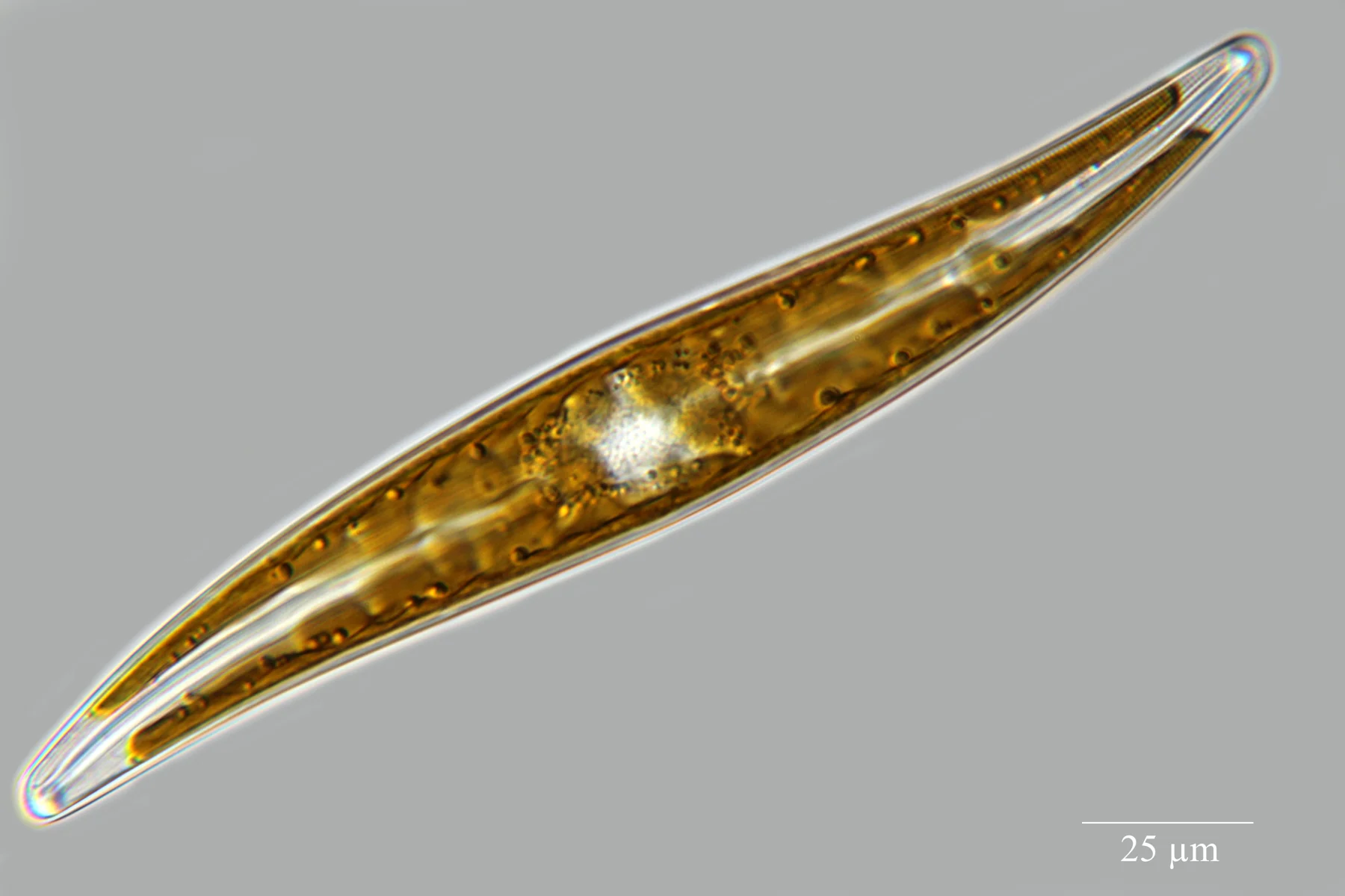
A long, golden Gyrosigma cell, slightly curved, enclosed in a transparent wall.
A living diatom photographed using differential interference contrast. Anatoly Mikhaltsov · Source · CC BY-SA 4.0
View the full photograph ↗

One cell

An alga inside a silica shell.

Gyrosigma acuminatum

Diatoms build their cell walls from silica, a material also used to make glass. The transparent shell reveals the golden-brown interior of this living cell.

LOOK HERE

Follow the long outline, curved into a gentle S. Inside are the chloroplasts that help the alga use light. The full photograph retains the author’s scale bar.

About this organism ↗

Photographs of different specimens, shown at different scales.

Tiny does not mean simple

A single cell can do all this.

Micrasterias uses light. Stentor feeds and can rebuild parts of its body. Through a microscope, an organism’s outline cannot tell you how many cells it contains.

One cell

In Micrasterias, the whole branching shape belongs to one cell.

A colony

In Volvox, many cells form a sphere together.

An animal

In a rotifer, cells form tissues and organs.

The rotifer and its apparent wheels

Why do cilia look
like turning wheels?

Cilia are fine filaments that bend one after another. Together, their movements look like rotation. The name “rotifer” comes from this impression of a turning wheel.

Follow the marked, thicker filament. Its base stays in the same place while the movement travels around the circle.

Lander University · Philodina ↗

FIXED BASES
A schematic movement, slowed down so you can follow it.

Now try without the labels

One cell, a colony
or an animal?

Five photographs. Look at the shape, choose an answer and see what you recognised.

Microscope photograph for identification
?

What are you looking at?

How to tell them apart

In Micrasterias, the whole branching shape belongs to one cell. In Volvox, many cells form a sphere together. In a rotifer, cells form tissues and organs.

Next time you pass a pond

What else might you find
in a pond?

Films on plants, clumps of algae and sediments can hold very different organisms. They do not all appear in the same sample. What you find depends on where you collect the water and when you look at it.

The Quekett Microscopical Club’s illustrated guide will help you recognise more of the life in a pond.

See the observation guide ↗

How we know

The photographs show real specimens. The explanations draw on the sources below; general descriptions do not replace species identification.

Photographs and colours

These photographs come from separate observations. Illumination and microscopy techniques affect how colours and outlines appear. Enlargement here brings the photograph closer; it does not represent a microscope’s optical magnification.

Images have been resized for the page and framed in circles in the explorer. The full photograph retains the author’s composition and scale bars. Photo credits and licences are listed with each specimen.