An experience about aging eyes
The face she can no longer see is yours
Look at a family visit through your grandmother's eyes, rebuilt from measurements of aging vision. The light dims, the small print escapes — and the face she turns towards dissolves into the spot her brain paints over.
Photograph: a grandmother holding her grandchild, Guangxi, China, 2010, by Star-trooper, CC BY 2.0, via Wikimedia Commons; cropped and resized.

The visit
She hears you before she sees you — the gate, your step on the path. Then she looks up, straight at you, and smiles.
This is what you see: her grandchild in her arms, every eyelash sharp. Keep this picture. You are about to watch it age.
Now step into her place. She is 78, and she turns towards the child’s face the way you turn towards this photograph.
What follows is that face through her eyes, rebuilt from measurements of aging human vision. The years first. Then the middle.
The years
At 45, the small print starts to escape: Hofstetter’s average formula gives a near point of about 20 centimetres — closer than that, and print dissolves.
Come on Sunday. Lunch at one, don’t be late. — Mama
By the mid-60s the world has gone a little dimmer and flatter. Her pupils open less wide than yours — about 0.04 millimetres narrower every year at the study’s lowest light level — and her lens lets less light through, least of all the blue.
The US National Institutes of Health put it plainly: people in their 60s need three times more light for comfortable reading than people in their 20s. Stand her lamp closer. It is not fussiness; it is physics.
And yet noon looks white to her too. Her lens has been yellowing for decades, and her brain has spent those decades rebalancing the colour over the years — resetting white the way a camera does.
After cataract surgery floods the eye with blue again, the neutral point shifts back over a number of months, without ever fully returning. That is a brain that refuses to see the world yellow. Remember this repair crew. You will meet them again.

The missing middle
She is 78 now. Watch the child’s face — the face she turns towards.
AMD itself is common: a global meta-analysis estimated some form of it in about one adult in twelve aged 45 to 85. Most of that was early disease.
The blind centre you are about to see is an advanced outcome, not what everyone with AMD experiences: in advanced disease, damage to the macula can produce a central blind zone. She is one woman whose disease has taken that form. Many, not all. This is her.
The middle is going. Not blurred — gone, and painted over with the surroundings, the way a restorer fills a hole in a fresco.
She would probably not describe a spot or a shadow. In a study of 153 people seen for low vision with this condition, 88% had a blind zone in both eyes; more than half of those had no idea it existed — even with dense ones up to 30 degrees across. Nearly half only ever noticed things ‘disappearing’.
This page is not a vision test. New changes in sight deserve an eye doctor.
Two views of the same eyes
Her brain rebalances the colour and paints the middle over. White is white to her — and nothing looks missing.


She turns towards you and smiles. The middle of you is gone — and she cannot see that it is gone.
Ordinary glasses cannot refocus this part: the loss is in the retina, and the brain paints over it. Low-vision aids and rehabilitation cannot restore the retina either — but they can help people use the vision that remains. That is the cruel fact this page is about.
Your brain too
You may not believe a brain can delete the middle of the world and tell no one. So here is yours doing it.
In 1991, Ramachandran and Gregory showed that a grey square on flickering static vanishes after about 10 seconds of steady staring — filled in by the surroundings. Stare, and watch your eyes lie to you.
Hold your eyes still
Cover one eye, stare at the centre dot, and keep your gaze as steadily as you can on the dot. Within about ten seconds the grey square may dissolve into the static as the moving surround fills its place.
Still image: with movement reduced, the square sits on static noise. Stare at the dot; it may fade and return.
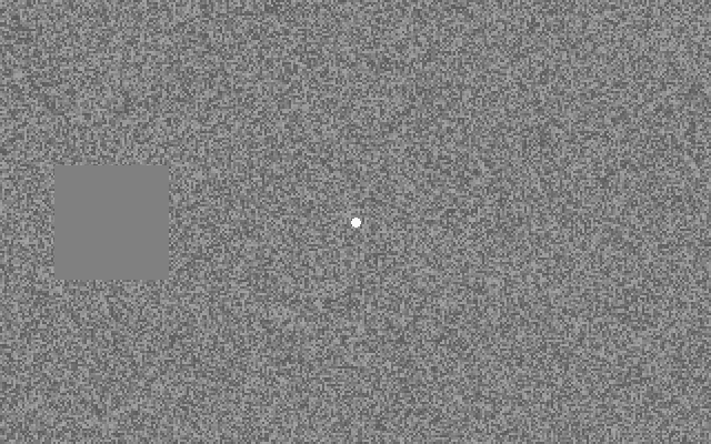
Yours is intact vision playing a trick; hers is damaged retina. The two are not biologically identical — but a separate study found the same kind of completion across real AMD blind zones: in both, a region can disappear without presenting itself as a black hole.
In this artificial-scotoma experiment, tiny eye movements help bring the square back: they falter, and it dissolves; they resume, and it returns. That finding comes from intact eyes — it does not tell us the same timing controls filling-in around an AMD blind zone.
Your photograph
See your own photograph as she does
Choose a photograph of your family. It stays on this device and disappears when you close the page.
On the interactive page, you can watch your own photograph age the same way.
This is the visit through her eyes: your face, the one she looks for first — present, loved, and half-painted.
She knows you are there. She hears you, she feels your hand, she turns toward your voice. What she cannot do is check your face against your voice. So give her everything else.
The next visit
Stand where the light falls on your face. She needs the light on what she looks at.
Come close enough to make your face easier to use, and ask what distance works best. Let her choose where to look: with central vision loss, the clearest view may be slightly off-centre. Say your name when you walk in, before she has to ask. None of this restores the middle. All of it reaches her around it.
Sit in the light, sit close, and say your name when you walk in.
These are your eyes again — the whole face present. One day they will be old too. Until then, spend them looking at her.
Sources and method
The photograph ages through illustrative mappings of measured declines, in healthy eyes: contrast from a 292-person study that found sensitivity falling 0.03 log units a decade (Peñaloza & Kwon 2025), blur from its acuity slope, dimming from the shrinking pupil (0.043 mm a year at the study’s lowest light level; Winn et al. 1994) and the densifying lens. These are renderings of numbers, not measurements of appearance.
The yellowing appears only in the capture view, and only at the measured group difference: lenses over 60 let through 40% less light at 420 nm than lenses of 40–59, grading to 18% less at 580 nm (Artigas et al. 2012, 32 lenses, measured outside the body). The perceived view stays white because the brain rebalances colour over decades: after cataract surgery the neutral point returns over a number of months but never fully (Delahunt et al. 2004, 4 patients). The capture tint is a schematic RGB mapping anchored to those measured spectral differences; it is not a colourimetric prediction of an older observer’s retinal image.
The dissolve is drawn at one possible extent of a central blind zone. Dense ones up to 30° across were found in referred patients (Fletcher et al. 2012), completion has been shown across 7° of radius (Zur & Ullman 2003), and no single typical size exists. The painted middle illustrates what patients report: binocular scotomas were found in 88% of the 153 patients, and 56% of that subgroup were totally unaware of them. Matching these numbers shows the build is faithful, not that the biology is certain.
The prevalence figures distinguish any disease from late disease: 8.69% any, 8.01% early and 0.37% late, mapped to ages 45–85 (Wong et al. 2014).
The scene keeps the missing region centred on the face to make the loss legible. People with established central vision loss often adopt an eccentric preferred retinal locus instead of the damaged fovea (Crossland et al. 2005: all 25 patients within 6 months), so real fixation strategies can differ.
The 20-centimetre near point at 45 is the average of Hofstetter's formula (18.5−0.30×age dioptres), quoted via a 2026 review — a formula, not measured individuals.
The staring panel recreates the Ramachandran & Gregory 1991 paradigm — a grey square on twinkling noise of equal mean luminance. Their square subtended 1.5°; ours is an uncalibrated screen recreation, not a reproduction at that visual angle. Tiny eye movements counteract the fading in this paradigm (Troncoso et al. 2008, building on Martinez-Conde et al. 2006). Not every reader will see it fade.
This page is not a vision test, and it predicts nothing about any one person's eyes: macular degeneration is probabilistic — many, not all. Low-vision aids and rehabilitation cannot restore the retina, but a systematic review of 35 studies reported benefits for acuity, reading and face recognition (Macnamara et al. 2023, certainty of evidence low-moderate). New changes in sight deserve an eye doctor.
- Peñaloza & Kwon (2025), Significant age-related visual declines but preserved binocular summation, Invest Ophthalmol Vis Sci 66(14):63 doi.org/10.1167/iovs.66.14.63
- Owsley, Sekuler & Siemsen (1983), Contrast sensitivity throughout adulthood, Vision Res 23:689–699 doi.org/10.1016/0042-6989(83)90210-9
- Artigas, Felipe, Navea, Fandiño & Artigas (2012), Spectral transmission of the human crystalline lens, Invest Ophthalmol Vis Sci 53:4076–4084 doi.org/10.1167/iovs.12-9471
- Pokorny, Smith & Lutze (1987), Aging of the human lens, Appl Opt 26:1437–1440 doi.org/10.1364/ao.26.001437
- Eto et al. (2020), A Purkinje image-based system for assessment of the human crystalline lens in vivo, Sci Rep doi.org/10.1038/s41598-020-73541-y
- van de Kraats & van Norren (2007), Optical density of the aging human ocular media, J Opt Soc Am A 24:1842–1857 doi.org/10.1364/josaa.24.001842
- Chrysanthopoulos, Pateras & Plakitsi (2026), Assessment and clinical classification of accommodative dysfunctions, Cureus 18:e105684 doi.org/10.7759/cureus.105684
- Winn, Whitaker, Elliott & Phillips (1994), Factors affecting light-adapted pupil size, Invest Ophthalmol Vis Sci 35:1132–1137 pubmed.ncbi.nlm.nih.gov/8125724/
- NIH News in Health, Your Aging Eyes, December 2017 (guidance) newsinhealth.nih.gov/2017/12/your-aging-eyes
- Delahunt, Webster, Ma & Werner (2004), Long-term renormalization of chromatic mechanisms following cataract surgery, Vis Neurosci 21:301–307 doi.org/10.1017/S0952523804213025
- Neitz, Carroll, Yamauchi, Neitz & Williams (2002), Color perception is mediated by a plastic neural mechanism, Neuron 35:783–792 doi.org/10.1016/S0896-6273(02)00818-8
- Wong et al. (2014), Global prevalence of age-related macular degeneration, Lancet Glob Health 2:e106–116 doi.org/10.1016/S2214-109X(13)70145-1
- Fletcher, Schuchard & Renninger (2012), Patient awareness of binocular central scotoma in age-related macular degeneration, Optom Vis Sci 89:1395–1398 doi.org/10.1097/OPX.0b013e318264cc77
- Zur & Ullman (2003), Filling-in of retinal scotomas, Vision Res 43:971–982 doi.org/10.1016/S0042-6989(03)00038-5
- Ramachandran & Gregory (1991), Perceptual filling in of artificially induced scotomas, Nature 350:699–702 doi.org/10.1038/350699a0
- Ramachandran (1992), Filling in the blind spot, Nature 356:115 doi.org/10.1038/356115a0
- Martinez-Conde, Macknik, Troncoso & Dyar (2006), Microsaccades counteract visual fading during fixation, Neuron 49:297–305 doi.org/10.1016/j.neuron.2005.11.033
- Troncoso, Macknik & Martinez-Conde (2008), Microsaccades counteract perceptual filling-in, J Vis 8(14):15 doi.org/10.1167/8.14.15
- Crossland, Culham, Kabanarou & Rubin (2005), Preferred retinal locus development in patients with macular disease, Ophthalmology 112:1579–1585 doi.org/10.1016/j.ophtha.2005.03.027
- Macnamara, Chen, Davies, Sloan & Loetscher (2023), Low vision devices for age-related macular degeneration: a systematic review, Disabil Rehabil Assist Technol 18:998–1010 pubmed.ncbi.nlm.nih.gov/34416116/
- The photograph: Red Yao grandma holding her grandchild, Longji Terrace Fields, Guangxi, China, 2010, by Star-trooper, CC BY 2.0, via Wikimedia Commons; cropped and resized commons.wikimedia.org/wiki/File:Red_Yao_Child_and_grandma,_Longj
.jpg)