Week 4 · the limb bud
The arm is just a bud; the hand has no shape yet. A single vessel, the brachial artery, enters the bud.
Clinical anatomy · the persistent median artery
Press gently on your wrist, where you'd take your pulse: underneath the skin, two arteries usually run there, the radial and the ulnar. In a recent cadaveric study, a third one was also present in 43% of limbs: the median artery, a vessel that normally regresses during embryonic development, well before birth. In some of the limbs where it stays, it runs through the carpal tunnel and can become the sole blood supply of a finger. A surgeon who isn't looking for it risks cutting it.
Source: Ellis, Thibault, Lencke and Hemric, "Prevalence and anatomical significance of the persistent median artery: A cadaveric study," PLOS One, 31 March 2025. See method and sources below.
The mechanism
In the fourth week of embryonic development, the arm is a small bud, and the hand doesn't have a hand's shape yet. The brachial artery, coming down from the shoulder, branches for the first time in the fifth week: one of those branches, the median artery, feeds the forming hand at this stage. The radial and ulnar arteries develop alongside it and gradually take over. By the eighth week, in most embryos, the median artery regresses. In the rest, it stays.
Week 4 · the limb bud
The arm is just a bud; the hand has no shape yet. A single vessel, the brachial artery, enters the bud.
The slider works with the keyboard and with touch. The page respects the system's reduced-motion setting; nothing is saved in the browser.
Adult anatomy
Of the 43 limbs in 100 where the median artery stays, most have the antebrachial type: in 28 of 100, the vessel ends somewhere in the forearm, before the wrist. In 15 of 100, though, it crosses the carpal tunnel and reaches the palm, where it can become the sole blood supply of a finger.
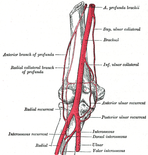
The hand is fed by the radial and ulnar arteries. The median artery no longer exists: it regressed during embryonic development.
What the measurements show
The researchers recorded whether the median artery was present and measured the diameter of the vessels around it. One result clears the usual bar for statistical significance; the other stays a trend.
A secondary finding, unrelated to the median artery
The cadavers also gave a comparison unrelated to the median artery: how similar the radial and ulnar arteries of the same person are, from one arm to the other. The answer surprised even the study's own authors.
In other words, if you know how thick someone's radial artery is on one arm, you can barely guess how thick it is on their other arm. Each limb seems to build its own map of blood vessels, largely on its own.
Why it matters to a surgeon
In an ordinary carpal tunnel release, the surgeon cuts the ligament covering the tunnel to free the compressed median nerve. If a palmar-type median artery is present and has become, further down in the palm, the sole blood supply of a finger, accidentally tying it off or cutting it can cut off circulation to that finger, with a risk of necrosis: tissue death from lack of blood.
A link between the palmar-type median artery and carpal tunnel syndrome has been proposed by other researchers, but causation remains unproven: it isn't known whether the artery contributes to compressing the nerve, or whether the two simply tend to occur together in the same tight space. Hand and wrist injuries are common: according to the authors, they account for 11% to 22% of all emergency-room visits in the United States.
Limits of the study
The 56 cadavers were between 62 and 100 years old at death, and information about vascular disease, prior trauma, and prior surgery to the hand and forearm was not available for any of them. The journal's editor flagged this limit explicitly. The authors also say this is the first study to measure the external diameter of the persistent median artery and its relationship to the surrounding vessels, which means the result is waiting on independent replication.
The overall rate of 43% sits close to a prior meta-analysis, by Solewski and colleagues, of 10,394 limbs, which reported 8.6% palmar-type and 34.0% antebrachial-type separately: added together, about 42.6%. But the split between the two types differs a great deal: this study found nearly double the palmar-type share found in that much larger meta-analysis. It isn't known whether the difference comes from the population studied, the dissection method, or chance, and the 15% figure remains an estimate from a single sample, awaiting confirmation from future studies.
Recap
The same story, in words and numbers: the development of the median artery, the three possible routes in an adult, and the full table of measurements.
The arm is a bud; the brachial artery branches for the first time, and the median artery is the main branch feeding the forming hand.
The radial and ulnar arteries develop in parallel and take over more and more of the hand's blood supply; the median artery gradually becomes surplus.
In most limbs (57 of 100, in the studied sample), the median artery regresses during embryonic development. In the rest (43 of 100), it stays open for the rest of life.
The median artery persists but ends somewhere in the forearm, before the carpal tunnel. It never reaches the hand.
The median artery crosses the carpal tunnel alongside the median nerve and continues into the palm, where it can become the sole blood supply of a finger.
A surgeon who cuts the carpal tunnel ligament without knowing about this vessel risks cutting off circulation to the finger it supplies.
| Measurement | Without / type | With / type | Significance |
|---|---|---|---|
| Persistent median artery (of 89 limbs) | — | 43% (38 limbs) | — |
| · antebrachial type | — | 28% (25 limbs) | — |
| · palmar type | — | 15% (13 limbs) | — |
| Superficial palmar arch | 2.02 mm | 2.47 mm (with palmar type) | p = 0.03 |
| Ulnar artery | 2.75 mm | 3.02 mm (with palmar type) | p = 0.08 (trend) |
| Left vs. right arm, radial artery | r² = 0.098 | — | |
| Left vs. right arm, ulnar artery | r² = 0.005 | — | |
Method and sources
This page starts from the study by Connor Ellis, Drew Thibault, Josh Lencke and Laurieanne D. Hemric (Liberty University College of Osteopathic Medicine), published in PLOS One on 31 March 2025. Every figure about prevalence, diameters and significance thresholds comes from that study. The comparison with the prior published rate comes from Solewski and colleagues' 2021 meta-analysis, cited directly in the article.
The page invents no clinical detail, surgical procedure or individual outcome. The routes shown in the interactive diagram follow the anatomical descriptions in the study; the weeks of development in the embryology figure follow the published timeline (formation around week five, usual regression by week eight). The diagrams are schematic drawings.
