
Seen in tissue
In post-mortem hippocampus, resident microglia thin out between about 50 and 75. A second population appears, with stronger inflammatory signals.
Brain immunity
In the of 40 donated brains, resident thin out from about age 50 to 75. Cells with a blood-lineage signature appear in the same window. suggests their origin. The evidence comes from tissue taken after death, and the study did not track memory.
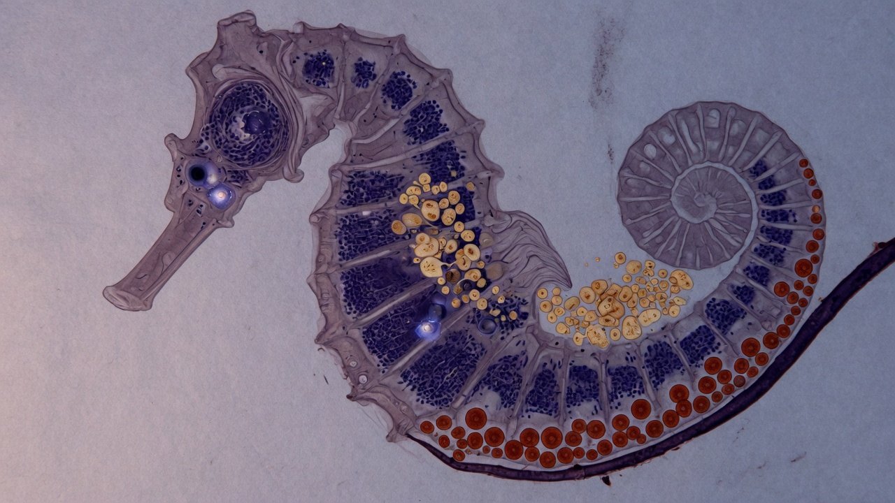
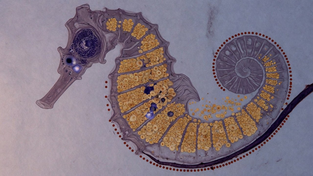
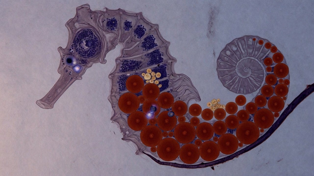
The name hippocampus comes from its resemblance to a seahorse. This is an editorial illustration, not a micrograph from the paper.
Move the age marker and watch the schematic change.
Between 50 and 75, the paper describes gradual replacement. All 40 published cases span ages 20–95.
The published 50–75 window is shown as a schematic. The mix is editorial; it is not a percentage taken from the paper's figures.
What the paper shows
In post-mortem hippocampus, resident microglia thin out between about 50 and 75. A second population appears, with stronger inflammatory signals.
The new population shares epigenetic traits with blood monocytes. The study did not trace these cells inside a living person.
The authors propose that the shift may contribute to chronic inflammation in neurodegenerative disease. The paper did not measure dementia in these donors.
We do not yet know whether the shift appears in living people over time or affects memory.
What the change looks like
are the brain's resident immune cells. They arise in the embryo and, in the classical model, renew themselves locally throughout life. In these 40 hippocampi, resident thin out after age 50.
Resident microglia arise in the embryo. They clear debris, monitor synapses and respond to danger signals. The paper finds fewer of them between about 50 and 75.
In their place, cells appear with a stronger inflammatory program and traits resembling blood . The team interprets the methylation pattern as a change in cell origin.
The public releases describe the donors as having no known neurological disease. That does not rule out pathology a clinician never detected.
Two ways to read one cell
Nathan Zemke, the first author, explains the distinction. Gene expression shows what a cell is doing now, while epigenetic signatures preserve clues about where it came from. The upper band mixes the points. Methylation separates them into two families below.
In gene expression, the groups mix.
Methylation shows two distinct patterns.
Gene expression tells us what a cell is doing today, but epigenetic signatures preserve information about where a cell came from.
Nathan R. Zemke, in the NIH recap of the paper
Who was studied
Each mark is one donor; positions are not real ages. Table S1 lists each case's age, sex and inclusion criteria. The Science PDF was unavailable at the date of this edition, so this grid does not copy the table.
How far the evidence goes
The paper reaches tissue and an origin suggested by methylation. No test or treatment has been evaluated for this change.
Researchers analyzed 40 hippocampi from donors aged 20 to 95, using tissue taken after death.
Methylation offers clues about cell origin. The study did not trace these cells inside a living person.
The new population carries a proinflammatory program. The study did not measure a cause of disease, so the link remains a hypothesis.
No longitudinal study has followed the same shift in living people.
There is no validated clinical biomarker for this change yet.
No therapy, supplement or protocol has been tested for this change.
What is worth asking
The paper describes the loss. The public releases do not say whether cells die, become exhausted or leave the tissue.
Methylation points toward a blood-like origin. A separate Stanford study, published later in Nature, describes immune cells entering from the periphery. Its design differs, and it does not confirm the 40 hippocampi in the Zemke paper.
The study measured the hippocampus. To generalize to the whole brain, researchers need to study other regions.
The public releases describe the donors as having no known neurological disease. The study did not track their memory over time.
Where the claim comes from
The Zemke paper and releases from the NIH and New York Genome Center support the cell findings. The diagrams are editorial and do not reproduce figures from the paper.
Concerns about memory, language or orientation belong with a clinician. The page does not assess individual risk and does not recommend tests or products.
Epigenetic and 3D genome reprogramming during the aging of the human hippocampus. Original research article. DOI 10.1126/science.adt8307. The PDF was unavailable at the date of this edition.
The NIH release gives the 50–75 window, the sample of 40 and the replacement reading of microglia.
Bing Ren's institutional release confirms the decline of embryonic microglia, the blood-like signature and the barrier finding.
This recap repeats the NIH release, including Zemke's quotation and the author list. It adds no independent measurement.
The 2010 reference for the embryonic, yolk-sac origin of microglia. The 2026 paper revisits this model in older adults.
A separate study of immune cells entering the aging brain from the body. It uses a different design and journal, and does not confirm the 40 hippocampi in the Zemke paper.
The open question
We still do not know what this change means for memory.