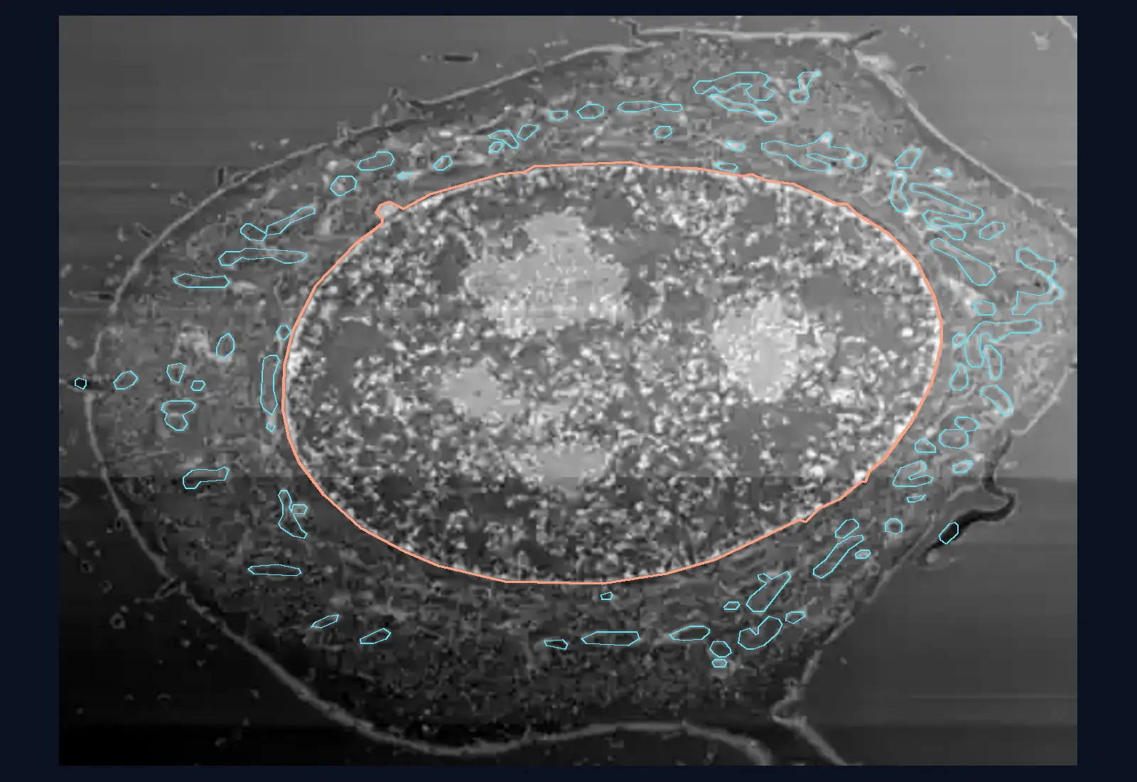
Chapter 3 · The cellLesson 3.5
Cell diversity and the surface area to volume ratio: why cells have different shapes and stay small
Why does a neuron have long extensions, why is a red blood cell flattened, and why is an elephant not made of one giant cell?
- Size
- from 5 nm to 100 µm
- Level
- cell
- Time
- a day in the life of a cell
Before you read: what do you think?
The walls of the small intestine are covered in folds, villi and microvilli. According to measurements from 2014, how large is the inner surface of an adult’s whole digestive tract?
See the answer
About 32 m², of which about 2 m² is in the large intestine. Old textbooks gave 260–300 m², but those estimates rested on incomplete data. Even so, villi and microvilli make the surface of the small intestine 60–120 times as large.
A motor neuron in the spinal cord can have an axon a metre long, from the base of the spine to the toes. A red blood cell measures only 7–8 µm and is flattened like a tablet hollowed out on both faces. Both are cells of the same person. The shape of each fits its work.
Animal cells: a shape for every job
The neuron receives and transmits signals. It has a cell body, with the nucleus, many branched dendrites, which receive messages from other neurons, and a single axon, which carries the signal to another cell, sometimes over a long distance.
The red blood cell (erythrocyte) carries oxygen. It is shaped like a biconcave disc, thick at the edges and very thin in the middle, and this shape gives it a large surface area compared with its volume, through which gases pass quickly. A mature red blood cell has no nucleus and no mitochondria, and it folds to squeeze through the narrowest capillaries.
The skeletal muscle fibre contracts. It is a very long cell with many nuclei, formed in the embryo when hundreds of cells fuse. In the sartorius muscle of the thigh it can reach 30 cm in length and 100 µm in thickness.
Epithelial cells cover surfaces and line organs. Those of the small intestine have microvilli on the side facing the inside of the gut: tiny extensions of the membrane that increase the surface through which substances are absorbed.
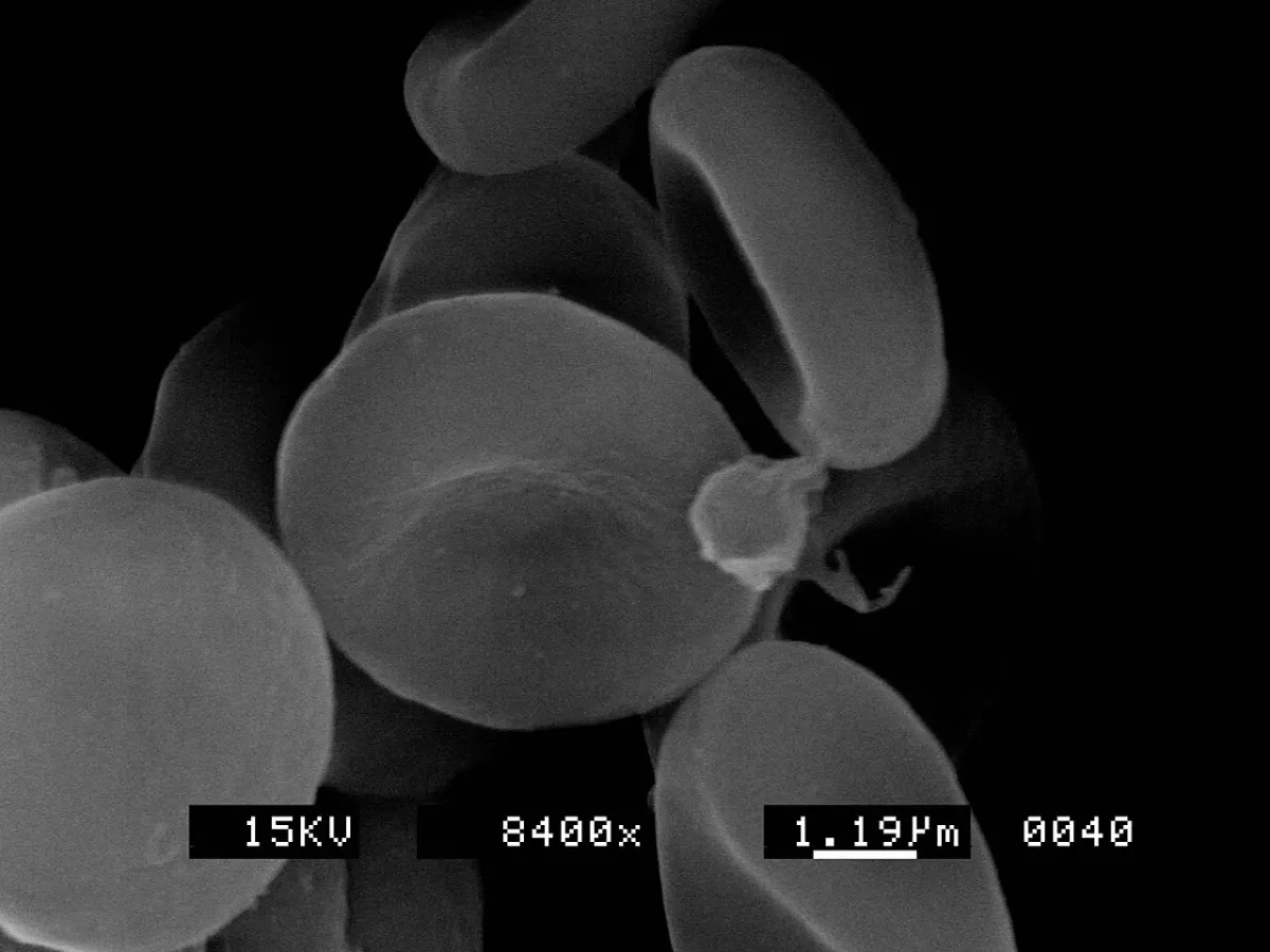
Photo: Scootdive, CC BY-SA 3.0, source. Cropped (top part) and resized.

Plant and fungal cells: shapes for water, gases and food
Root hairs are extensions of cells in the root epidermis. They greatly increase the surface through which the root absorbs water and mineral salts.
Guard cells are pairs of cells that surround each stoma in the leaf epidermis. They open and close the stoma, the opening through which the leaf exchanges gases with the air.
Xylem vessels are tubes made of cells that are dead at maturity, with thick walls hardened with lignin. The end walls of the cells are perforated, so cells placed end to end form the tubes through which water rises. Phloem sieve tubes, which carry phloem sap, are made of living cells without a nucleus, helped by neighbouring companion cells.
In fungi, the body is made of hyphae, thin branching filaments that together form the mycelium. In most fungi the hyphae are divided by cross walls; in others, such as bread mould, they are continuous tubes with many nuclei. Fungi secrete their enzymes outside the body and absorb the broken-down substances through the large surface of the mycelium.
Why cells stay small: surface area and volume
Everything that enters or leaves a cell passes through the membrane, that is, through its surface. What uses up food and oxygen is the volume, the inside of the cell. When a cell grows and keeps its shape, its volume increases faster than its surface area.
For a cube with side a, the surface area is S = 6a² and the volume is V = a³. The surface area to volume ratio is S/V = 6/a. If the side doubles, the surface area grows fourfold, the volume eightfold, and the ratio halves. A large cell has less membrane, for each cubic micrometre inside it, through which to take in substances and get rid of waste.
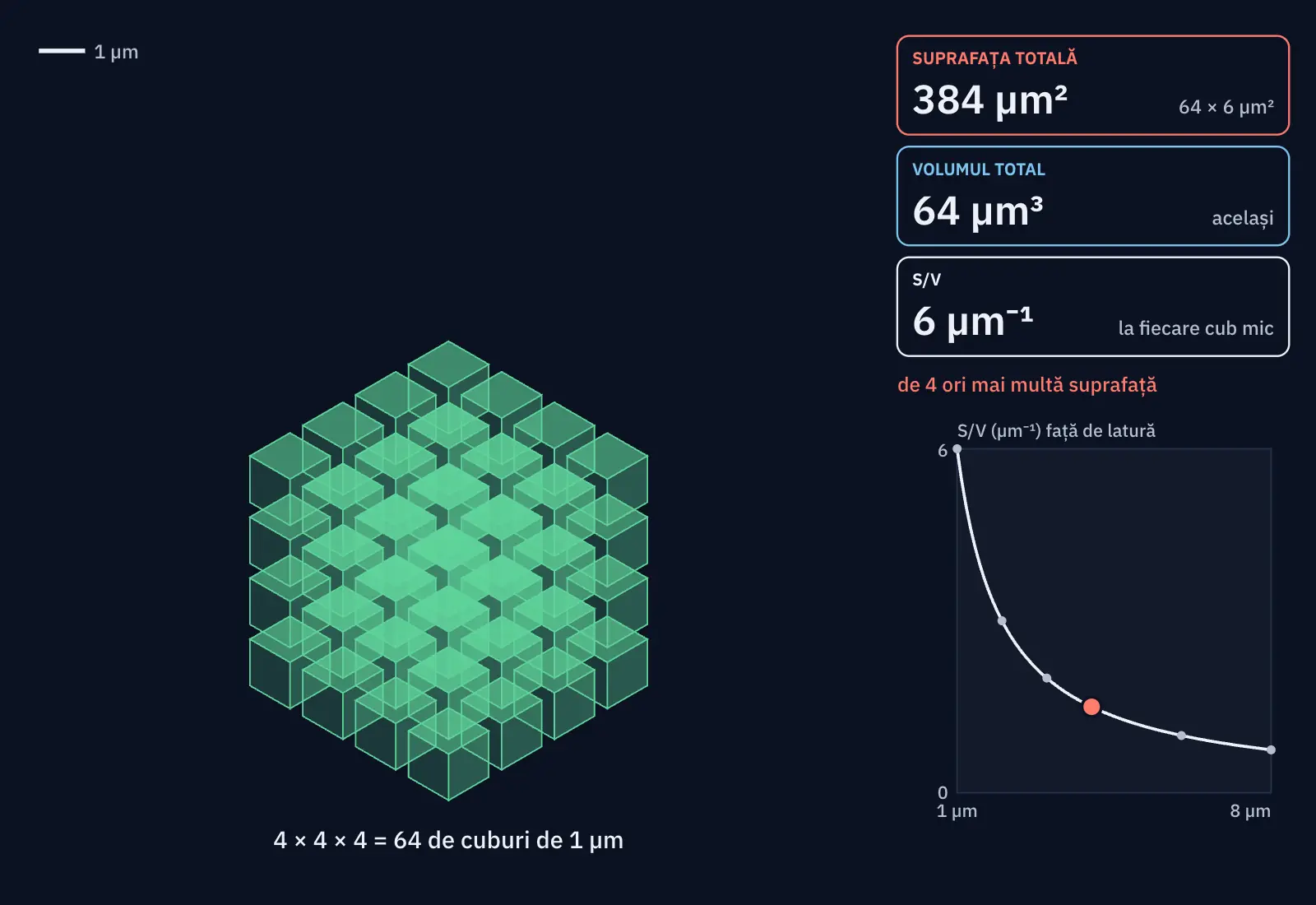
| Side a | Surface area S | Volume V | S/V | Cut into cubes of 1 µm: total surface area |
|---|---|---|---|---|
| 1 µm | 6 µm² | 1 µm³ | 6 µm⁻¹ | 6 µm² (the same) |
| 2 µm | 24 µm² | 8 µm³ | 3 µm⁻¹ | 48 µm² (2 times as much) |
| 3 µm | 54 µm² | 27 µm³ | 2 µm⁻¹ | 162 µm² (3 times as much) |
| 4 µm | 96 µm² | 64 µm³ | 1.5 µm⁻¹ | 384 µm² (4 times as much) |
| 6 µm | 216 µm² | 216 µm³ | 1 µm⁻¹ | 1,296 µm² (6 times as much) |
| 8 µm | 384 µm² | 512 µm³ | 0.75 µm⁻¹ | 3,072 µm² (8 times as much) |
| Model time τ | Amount on the left | Amount on the right | Concentration on the left | Concentration on the right |
|---|---|---|---|---|
| 0 | 8 | 0 | 8 | 0 |
| 0.5 | 5.08 | 2.92 | 5.08 | 0.97 |
| 1 | 3.58 | 4.42 | 3.58 | 1.47 |
| 2 | 2.42 | 5.58 | 2.42 | 1.86 |
| 3 | 2.11 | 5.89 | 2.11 | 1.96 |
| 5 | 2.01 | 5.99 | 2.01 | 2 |
| equilibrium | 2 | 6 | 2 | 2 |
The compartments have volumes 1 and 3; at the start the amount 8 is all on the left. The total stays 8. At equilibrium the concentrations are equal, 2, and the amounts differ, 2 and 6, because the volumes differ. The distance, as an estimate: with D = 2, a root-mean-square displacement of 2 along one direction needs the time 1, and one of 4 needs the time 4: double the distance, four times the time.
Calculation. Scale drawings of idealised shapes: cubes without folds, and two perfectly mixed compartments with fixed volumes. S = 6a², V = a³ and S/V = 6/a; the ratio has the unit 1/length. In part B the volumes, the amounts and the time are in model units, and the model time τ has no seconds; the starting case (volumes 1 and 3, amount 8 on the left) is a worked example. The particles are only a drawing.
How cells and organisms increase their exchange surface
Large organisms are made of many small cells, each with a large surface area to volume ratio. Where exchanges are intense, cells are flattened, elongated or have extensions: the disc of the red blood cell, root hairs, microvilli, hyphae.
At the level of organs, the surface is increased by folds. According to measurements published in 2014, the inner surface of an adult’s digestive tract is about 32 m², and villi and microvilli make the surface of the small intestine 60–120 times as large as that of a smooth tube.
Worked example
Three cube-shaped cells
Real cells are not cube-shaped; the cube just makes the calculation easy. Compare three cubes with sides of 1 µm, 2 µm and 4 µm.
Side 1 µm: S = 6 × 1² = 6 µm², V = 1³ = 1 µm³, S/V = 6 µm⁻¹. Side 2 µm: S = 6 × 4 = 24 µm², V = 8 µm³, S/V = 3 µm⁻¹. Side 4 µm: S = 6 × 16 = 96 µm², V = 64 µm³, S/V = 1.5 µm⁻¹. Each time the side doubles, the ratio halves.
If you split the 4 µm cube into eight 2 µm cubes, the total volume stays 64 µm³, but the total surface area becomes 8 × 24 = 192 µm², twice as much. Dividing into small cells gives more membrane for the same volume.
At home you can see the same thing with a potato: cut a cube with 1 cm sides and one with 3 cm sides and leave them for an hour in beetroot juice diluted with water. The colour soaks in to about the same depth in both, but when you cut them in half, the small cube is coloured almost all the way through, while the large one has a white core.
In Romania
Gheorghe Marinescu and the nerve cell
Gheorghe Marinescu was born in Bucharest in 1863 and studied neurology in Paris with Jean-Martin Charcot, at the Salpêtrière hospital. Back home, he headed the University of Bucharest’s clinical neurology department at Colentina Hospital for 41 years, and he is regarded as the founder of the Romanian school of neurology.
In 1909, in Paris, he published a two-volume treatise, La cellule nerveuse (“The Nerve Cell”), with a preface by the Spanish histologist Santiago Ramón y Cajal. The first volume deals with the structure of the cell in general, the second with the neuron. Marinescu studied the changes in the neuron’s cell body after the axon is injured and described the fine fibres inside the nerve cell.
Between 1898 and 1901, also in Bucharest, he filmed patients with walking disorders, some of the world’s first scientific films.
In short
- The shape of cells is related to their function: the neuron has dendrites and a long axon, the red blood cell is a biconcave disc with no nucleus, the muscle fibre is long and has many nuclei.
- In plants, root hairs increase the absorbing surface, guard cells open and close the stomata, and xylem vessels are tubes of dead cells with lignified walls.
- In fungi, hyphae form the mycelium and absorb food through its large surface.
- For a cube with side a, S = 6a², V = a³ and S/V = 6/a; as a cell grows, its surface area to volume ratio falls.
- Cells stay small and are flattened, elongated or have extensions (microvilli, root hairs) because exchanges with the surroundings happen through the surface.
Common mistakes
WrongIf you double the side of a cell, its volume doubles too.
RightVolume increases with the cube of the side, so eightfold; the surface area grows only fourfold, and the surface area to volume ratio falls by half.
WrongA cell chooses the shape it needs.
RightA cell’s shape results from its genetic information and its development, and shapes suited to a job have been kept through evolution. The cell chooses nothing.
WrongA bigger cell exchanges substances better with its surroundings, because it has a bigger membrane.
RightIts membrane is bigger in total, but the volume it has to serve grows faster. Each cubic micrometre of the inside is left with less membrane.
Check yourself
Six questions. For the first five you see the explanation at once, with the paragraph of the lesson that supports it; for the written ones you compare with a good answer.
What does a red blood cell gain from its biconcave disc shape?
Answer
b) A large surface area compared with its volume, for gas exchange. The flattened, hollowed shape brings the whole inside close to the membrane and increases the surface area compared with the volume. A mature red blood cell has no nucleus or mitochondria and no wall.
The side of a cube increases from 2 µm to 4 µm. What happens to its volume?
Answer
c) It grows eightfold. V = a³, so (2a)³ = 8a³. The volume goes from 8 µm³ to 64 µm³.
Which description fits xylem vessels?
Answer
b) Tubes of cells that are dead at maturity, with lignified walls, placed end to end. Xylem vessels carry water through tubes made of dead cells. Living cells without a nucleus form the phloem sieve tubes, and the other options describe guard cells and root hairs.
What is the surface area to volume ratio of a cube with a side of 5 µm, in µm⁻¹?
µm⁻¹Answer
1.2 µm⁻¹. S/V = 6/a = 6 ÷ 5 = 1.2 µm⁻¹. Check: S = 150 µm², V = 125 µm³, 150 ÷ 125 = 1.2.
Choose the group each specialised cell belongs to.
Answer
Plant cell: Root hair; Guard cell; Xylem vessel cell.
Animal cell: Neuron; Red blood cell; Skeletal muscle fibre.
Fungal cell: Hypha.
Root hairs, guard cells and xylem vessels belong to plants; the neuron, the red blood cell and the muscle fibre to animals; hyphae are the filaments that make up the body of fungi.
An amoeba is a single, roughly round cell. Explain why it cannot keep growing for ever while keeping its shape, and what usually happens when it reaches a certain size.
A good answer
As the amoeba grows, its volume increases with the cube of its size and its surface area only with the square, so the surface area to volume ratio falls. The membrane, now relatively smaller, cannot let in enough food and oxygen, or let out enough waste, for the whole inside. At a certain size the cell usually divides into two smaller cells, each with a larger surface area to volume ratio.
A complete answer says that volume increases faster than surface area, links exchanges to the membrane (surface) and consumption to the volume, and names a solution: division or a change of shape.
Practical work for this lesson
- A bigger cube: what does it gain and what does it lose? – Geometric modelling, 35–40 minutes (CS)
Terms
- surface area to volume ratio
- The surface area of a cell divided by its volume; it falls as the cell grows while keeping its shape.
- neuron
- A nerve cell, with a cell body, dendrites and an axon, specialised in receiving and transmitting signals.
- axon
- The single extension of a neuron that carries the signal to another cell.
- microvilli
- Tiny extensions of the membrane of some epithelial cells that increase the absorbing surface.
- root hair
- An extension of a cell in the root epidermis that increases the surface for absorbing water and mineral salts.
- guard cells
- The pair of cells that surrounds a stoma and opens or closes it.
- hypha
- A thin filament of which the body of a fungus is made; many hyphae form the mycelium.
Sources and link to the programme
- OpenStax – Biology 2e, 35.1 Neurons and Glial Cells – the axon of a human motor neuron can be a metre long, from the base of the spine to the toes; dendrites receive messages; the diversity of neuron shapes
- OpenStax – Anatomy and Physiology 2e, 12.2 Nervous Tissue – the cell body, the dendrites, a single axon, the axon terminals
- OpenStax – Anatomy and Physiology 2e, 18.3 Erythrocytes – the 7–8 µm diameter; biconcave disc with a large surface area compared with its volume for gas exchange; no nucleus and no mitochondria; folds in capillaries
- OpenStax – Anatomy and Physiology 2e, 10.2 Skeletal Muscle – multinucleate fibres formed by the fusion of up to hundreds of myoblasts; diameter up to 100 µm and length up to 30 cm in the sartorius
- OpenStax – Biology 2e, 30.3 Roots – root hairs are extensions of root epidermal cells and increase the surface for absorbing water and minerals
- OpenStax – Biology 2e, 30.2 Stems – xylem vessel elements dead at maturity, with lignified walls and perforation plates at the ends; living sieve-tube elements without a nucleus, with companion cells; guard cells open and close the stoma
- OpenStax – Biology 2e, 24.1 Characteristics of Fungi – hyphae and mycelium; septate hyphae in most fungi, coenocytic in bread mould; absorption through the large surface of the mycelium after enzymes are secreted
- OpenStax – Biology 2e, 4.2 Prokaryotic Cells – as a cell grows, its surface area to volume ratio falls
- Helander, Fändriks – Surface area of the digestive tract – revisited, Scand J Gastroenterol 49(6):681–689, 2014 (PubMed) – the inner surface of the digestive tract, on average about 32 m², of which about 2 m² in the large intestine; villi and microvilli make the surface of the small intestine 60–120 times as large; the old textbook values, 260–300 m²
- Opris et al. – George Marinesco in the Constellation of Modern Neuroscience, Frontiers in Neuroscience 11:726, 2017 (PMC) – born in Bucharest in 1863; studied with Charcot at the Salpêtrière; chair of clinical neurology at Colentina for 41 years; the Romanian school of neurology; La cellule nerveuse, Paris, 1909, two volumes, preface by Ramón y Cajal; chromatolysis and neurofibrils; the scientific films of 1898–1901
From the programme: “Cell diversity related to functional specialisation in plant, animal and fungal cells”; “The importance of cell volumes and surface areas in the exchange of substances between the organism and its environment” (common core). Specific competences addressed: 1.1, 1.2, 2.2.