Chapter 3 · Inside the cell
What does one of your cells have that a bacterium does not?
- Size
- from 5 nm to 100 µm
- Level
- cell
- Time
- a day in the life of a cell
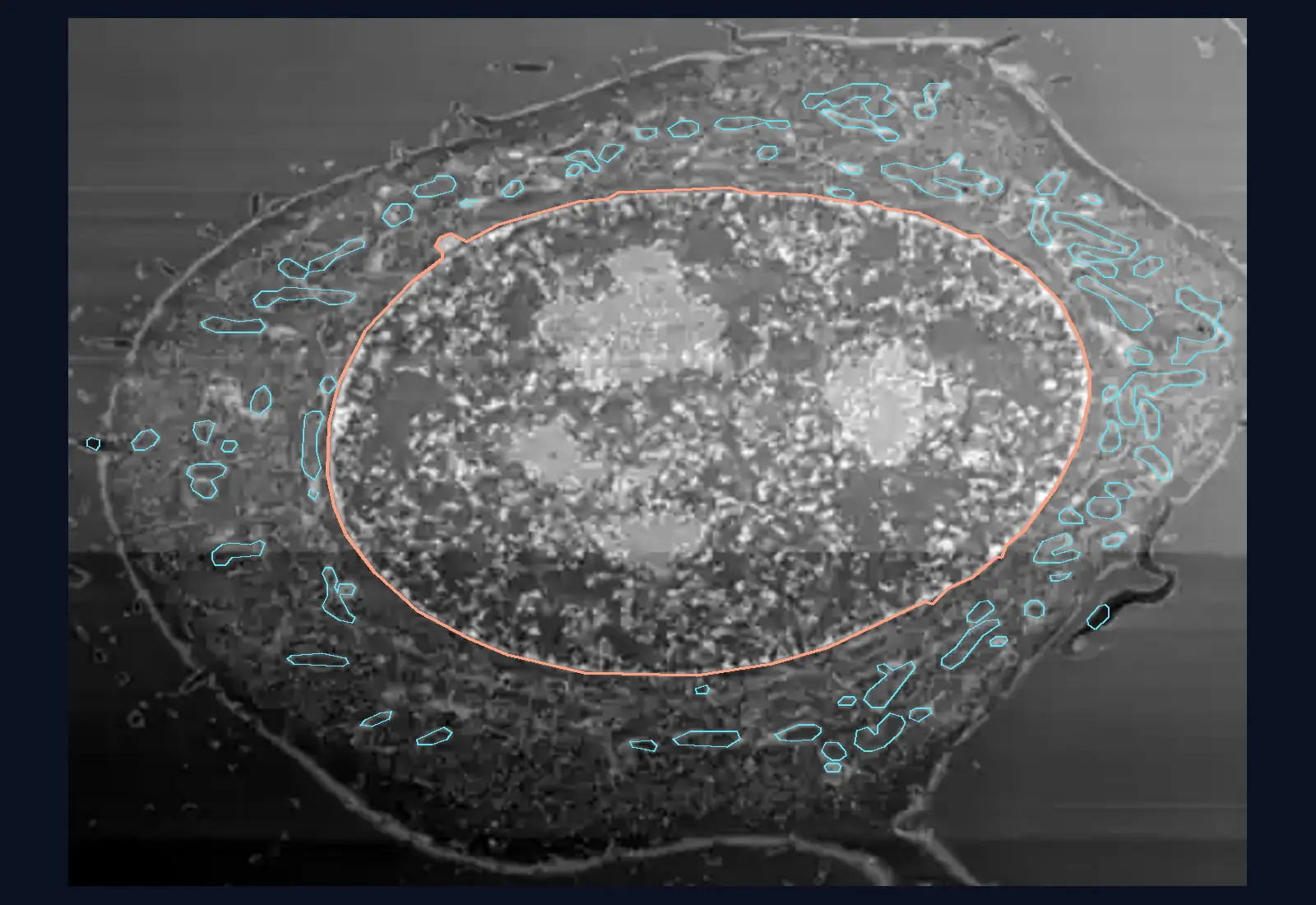
A human cell, grown in the laboratory, cut into 60 parallel planes from bottom to top. Each plane comes from the images of an electron microscope. The outlines of the nucleus, the mitochondria and the Golgi apparatus are drawn by a program, not by hand.
A cell cut into 60 slices
The cell is a HeLa cell, a human cancer cell grown in the laboratory since 1951, from the tumour of a patient at Johns Hopkins Hospital. An electron microscope fitted with an ion beam removed a layer of 5.24 nm from it, one after another, and photographed the face that remained each time. The images stacked on top of one another form a volume.
The planes shown here cut this volume from bottom to top, 91 nm apart. The nucleus is the largest structure of the cell.
In how many of the 60 slices do you think the nucleus appears?
The nucleus appears in 53 of the 60 slices
The scanned volume is 6.4 µm tall, and the nucleus alone is 5.0 µm thick. In this volume, the nucleus takes up almost the whole height. The outlines of the whole cell are not calculated, so there is no way to say what share of the cell’s height the nucleus is. In the small image at the top, the lines show where the plane lies relative to the nucleus.
In slice 24, the widest, the nucleus measures 21.2 × 13.6 µm. Its volume is 731 µm³, calculated from the automatic segmentation.
In slices 4–9, near the base, the nucleus appears in two or three pieces: the plane passes through hollows in its surface.
A mitochondrion appears as a dot or as a worm, depending on how it is cut
In slice 24, the program outlined 73 mitochondrial profiles: 18 almost round, 29 elongated, at least two and a half times longer than wide, and 26 in between. A profile is the shape a mitochondrion leaves in the plane of the cut.
The 73 profiles belong to 59 separate mitochondria, so some are cut twice or more. The mitochondria of the textbook, drawn as beans, are in fact cuts through long, branching structures.
In this volume, slice 14 has 116 profiles, and slice 45 only 6: above the nucleus only a thin layer of cytoplasm remains.
The Golgi apparatus sits in a hollow of the nucleus
The dotted yellow outlines show where the program found the Golgi apparatus, the stack of sacs that packages proteins. It is largest in slice 6, at 16 µm², between the pieces of the nucleus. It appears in slices 2–11.
The endoplasmic reticulum can be seen in the same slices, as thin, coiled membranes among the mitochondria. Its outlines are not drawn here, because they are too numerous and too tangled for a small screen.
The nucleus is 11 times longer than a bacterium
The silhouette in the corner is an Escherichia coli bacterium, drawn to the same scale: about 2 × 0.5 µm. The nucleus in the image is 21.2 µm long, almost 11 bacteria end to end.
It is a scale drawing, not a micrograph. The bacterium has a membrane and a cell wall, and its DNA lies in the cytoplasm, with no membrane-bound nucleus: that is why the silhouette has no nucleus drawn in it. The real width varies between 0.25 and 1.0 µm, depending on how the cell grows.
What is missing from the image: the wall and the chloroplasts
The HeLa cell is an animal cell: it has no cell wall and no chloroplasts. The image beside it is a chloroplast from a bean leaf, under the electron microscope. Its frame is 4.5 µm wide, 7.3 times narrower than the frame of the HeLa cell.
The chloroplast has a double membrane and flattened sacs, the thylakoids, visible as bands. The green is colour added by the author: an electron microscope has no colour. The cell wall of a plant surrounds the plasma membrane from outside and is not visible in any slice of the HeLa cell.
A root cell is still a plant cell, although it has no chloroplasts. The 60 slices show a single cell, grown flattened in culture, not a cell from a tissue.
Now you explain
A classmate says: “Mitochondria are little beans, like in the textbook.” What would you show them in the 60 slices?
Compare with an answer
The textbook drawing shows a cut through the middle of a mitochondrion. In a real cell, mitochondria are long, branching structures, and a plane cuts them in different directions: some profiles come out round, others elongated, and one mitochondrion can show up in several profiles. In slice 24, 73 profiles come from 59 separate mitochondria.
The lesson on cell organelles, with the worked example and questions