Chapter 3 · The cellLesson 3.1
The cell, a living structure: why viruses are not considered alive
A flu virus has genetic material and multiplies. Why do biologists say it is not alive?
- Size
- from 5 nm to 100 µm
- Level
- cell
- Time
- a day in the life of a cell
Before you read: what do you think?
To make flu vaccine, factories need large amounts of flu virus. What do they usually grow it in?
See the answer
Fertilised hens’ eggs. A virus can multiply only in living cells, and a fertilised egg has living cells. The method has been used for more than 70 years and is still the most common way of producing flu vaccine.
For more than 70 years, flu vaccine has been made with fertilised hens’ eggs: the virus is injected into the egg and left for a few days to multiply. Put the same virus in a flask of nutrient broth, where bacteria grow without trouble, and it would not multiply at all. The difference between the egg and the flask is the living cell.
What everything alive has
A salt crystal grows in a solution, and a flame consumes fuel and spreads. Neither is alive, because a single property is not enough. Biologists recognise life by a set of characteristics of living things that occur together: cellular organisation, metabolism, growth and development, reproduction, response to stimuli (excitability), homeostasis and adaptation to the environment.
Metabolism is the sum of the chemical reactions by which an organism takes energy and substances from its surroundings, transforms them and gets rid of what it does not use. Excitability is the ability to respond to stimuli: a plant bends towards light, a bacterium moves towards food. Homeostasis is keeping the internal environment relatively constant, for example the amount of water and salts in a cell.
The smallest level at which all these characteristics are found is the cell. That is why the cell is called the structural and functional unit of life: below it there are molecules, but no life.
Cell theory, built over almost 200 years
In 1665, Robert Hooke looked at a thin slice of cork through a lens and called the little compartments he saw “cells”. They were in fact the walls of dead cells. In 1838, the botanist Matthias Schleiden described plants as made of cells, and in 1839 the zoologist Theodor Schwann showed that animal tissues are made of cells too.
That left the question of where new cells come from. In 1852, Robert Remak published evidence that a new cell forms when an existing cell divides. Rudolf Virchow made the idea famous in 1855 with the Latin formula omnis cellula a cellula, “every cell comes from a cell”.
Cell theory comes down to three statements: all organisms are made of one or more cells; the cell is the basic unit of structure and function in organisms; every new cell comes from an existing cell.
How a virus is built
A complete virus particle is called a virion. At its centre is a nucleic acid, either DNA or RNA, wrapped in a capsid, a protein coat made of subunits called capsomeres. Some viruses, such as the flu virus and HIV, also have a lipid envelope, taken from the membrane of the cell they infected.
Most viruses measure between 20 and 250 nanometres. The light microscope in the school lab cannot separate details smaller than about 200 nanometres, so most viruses cannot be seen with it. The structure of viruses could be observed properly only after the electron microscope appeared, at the end of the 1930s.
A few examples you often come across: the flu virus and SARS-CoV-2, which causes COVID-19, have RNA and a lipid envelope; HIV also has RNA; bacteriophages are viruses that infect bacteria, and bacteriophage T4 keeps its DNA in a protein “head” and has a tail it uses to attach to the bacterium.
Why a virus is not considered alive
A virus is not a cell: it has no plasma membrane of its own, no cytoplasm, no ribosomes and no organelles. It has no metabolism of its own, produces no energy and cannot make its own proteins. Outside a cell, a virion is just a particle that may reach another cell, carried by air, by water or by contact.
To multiply, a virus has to get inside a suitable cell. The virion attaches to the surface of the cell, and the viral nucleic acid gets inside it. In many viruses the whole particle enters and then releases its nucleic acid; bacteriophages such as T4 inject only their nucleic acid into the bacterium, and the capsid stays outside. The cell, with its ribosomes and its energy, copies the viral nucleic acid and makes the virus’s proteins. These parts assemble into new virions, which leave the cell and infect other cells. That is why viruses are obligate intracellular parasites.
Viruses do have genetic material, though, and their populations change over time through mutations. For this reason some biologists call them entities “on the border of life”. The answer expected in class rests on cell theory: life starts at the cell, and a virus is not made of cells.
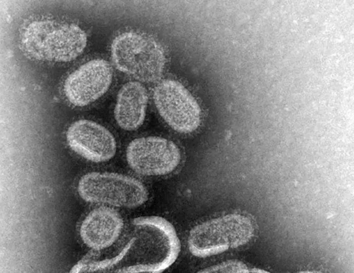
Photo: Cynthia Goldsmith; CDC / Dr. Terrence Tumpey, Public domain, source. Cropped (top part).

Worked example
Why a bacterium shows up under the school microscope and a virus does not
A rod-shaped bacterium is 2 µm long, and a flu virion is 100 nm across. The numbers are chosen for the exercise, within the usual ranges for each.
Step 1: put the lengths in the same unit. 1 µm = 1,000 nm, so 2 µm = 2,000 nm.
Step 2: compare. 2,000 ÷ 100 = 20, so the bacterium is 20 times longer than the virion.
Step 3: compare with the limit of the light microscope, about 200 nm. The bacterium, at 2,000 nm, is 10 times over the limit and appears as a small rod. The virion, at 100 nm, is below the limit and cannot be seen as a separate object.
In Romania
Victor Babeș and the virus he could not see
In 1888, the Romanian bacteriologist Victor Babeș opened the world’s second rabies vaccination centre in Bucharest, after the one Louis Pasteur had set up in Paris. Rabies is caused by a virus, but in those years nobody had seen a virus: the electron microscope would arrive only some 50 years later.
Babeș worked with what he could observe. In the nerve cells of rabid animals he described inclusions typical of the disease, rediscovered in 1903 by the Italian Adelchi Negri and known today as Babeș–Negri bodies. He also showed that heating the rabies virus to 58 °C for 2–14 minutes weakens it in a controlled way.
The institute of bacteriology and pathology founded in Bucharest in 1887 and run by Babeș bears his name today: the “Victor Babeș” National Institute of Pathology.
In short
- The characteristics of living things (cellular organisation, metabolism, growth and development, reproduction, excitability, homeostasis, adaptation) occur together, and the smallest level at which all of them are found is the cell.
- Cell theory says that all organisms are made of cells, that the cell is the unit of structure and function, and that every cell comes from an existing cell (Hooke 1665, Schleiden 1838, Schwann 1839, Remak and Virchow in the 1850s).
- A virion is made of a nucleic acid, DNA or RNA, and a protein capsid; some viruses also have a lipid envelope.
- Viruses have no cell, no ribosomes and no metabolism of their own, and multiply only in living cells, which is why they are obligate intracellular parasites.
- Most viruses measure 20–250 nm and cannot be seen with the light microscope; examples: the flu virus, HIV, SARS-CoV-2, bacteriophages.
Common mistakes
WrongA virus is a very small bacterium.
RightA bacterium is a cell, with a membrane, cytoplasm and ribosomes. A virus has no cell: it is a nucleic acid inside a capsid and multiplies only inside a cell.
WrongViruses are alive because they have DNA or RNA.
RightGenetic material is not enough. A virus has no metabolism of its own and cannot make its proteins outside a cell, and cell theory ties life to the cell.
WrongA crystal that grows is alive.
RightA crystal only adds more of the same material to its surface. It has no cells, no metabolism, no reproduction and no homeostasis.
Check yourself
Six questions. For the first five you see the explanation at once, with the paragraph of the lesson that supports it; for the written ones you compare with a good answer.
What does every cell have that a virus lacks?
Answer
b) Ribosomes. A virus has nucleic acid and has proteins in its capsid, and its genetic material undergoes mutations. It has no ribosomes, though, so it uses the ribosomes of the infected cell.
Which statement is part of cell theory?
Answer
d) Every new cell comes from an existing cell. Remak provided the evidence, and Virchow made the formula “every cell comes from a cell” famous. Viruses are not cells, and plants are made of cells too.
Why is flu virus for vaccines grown in fertilised eggs and not in a flask of nutrient broth?
Answer
c) Because the virus needs living cells to multiply. The virus uses the ribosomes and energy of the host cell. Nutrient broth feeds cells, bacteria for example, but contains no cells in which the virus could multiply.
The school light microscope cannot separate details smaller than about 200 nm. A virion measures 50 nm. How many times smaller than this limit is the virion?
timesAnswer
4 times. 200 ÷ 50 = 4. The virion is four times below the limit of the light microscope, so it cannot be seen as a separate object.
Put the stages of a virus multiplying inside a cell in order.
Answer
The correct order:
- The virion attaches to the surface of the cell
- The viral nucleic acid gets inside the cell
- The cell copies the viral nucleic acid and makes the virus’s proteins
- These parts assemble into new virions
- The new virions leave the cell and infect other cells
Unless the virion attaches to the cell, the viral nucleic acid cannot get in, and the cell copies the viral nucleic acid and makes the viral proteins on its ribosomes before the parts are assembled and released.
A classmate says: “Bacteriophages are bacteria, because you always find them next to bacteria.” Explain why this is wrong and what bacteriophages do to bacteria.
A good answer
Bacteriophages are viruses: they consist only of nucleic acid inside a protein capsid and have no cell, no ribosomes and no metabolism of their own. They are found next to bacteria because they infect them: they attach to a bacterium and insert their nucleic acid, and the bacterium makes new virions, which leave and infect other bacteria.
A complete answer says that a bacteriophage is a virus, names its structure (nucleic acid and capsid), shows that it lacks cellular organisation and explains that it depends on the bacterial cell to multiply.
Terms
- characteristics of living things
- The features that occur together in living organisms: cellular organisation, metabolism, growth and development, reproduction, excitability, homeostasis, adaptation.
- metabolism
- The sum of the chemical reactions by which an organism takes energy and substances from its surroundings, transforms them and gets rid of what it does not use.
- homeostasis
- Keeping the internal environment of a cell or an organism relatively constant.
- cell theory
- The theory that all organisms are made of cells, that the cell is the unit of structure and function, and that every cell comes from an existing cell.
- virion
- A complete virus particle, made of nucleic acid and a capsid, sometimes also a lipid envelope.
- capsid
- The protein coat that protects the nucleic acid of a virus.
- bacteriophage
- A virus that infects bacteria.
- obligate intracellular parasite
- An agent that can multiply only inside a living cell, using the cell’s components.
Sources and link to the programme
- CDC – How Influenza (Flu) Vaccines Are Made – the virus is injected into fertilised hens’ eggs and incubated for a few days; the method has been used for more than 70 years and is the most common one
- OpenStax – Biology 2e, 1.2 Themes and Concepts of Biology – the properties of life (order, response to stimuli, reproduction, adaptation, growth and development, regulation and homeostasis, energy processing); viruses are not made of cells and are not considered alive
- OpenStax – Microbiology, 3.2 Foundations of Modern Cell Theory – Hooke 1665, Schleiden 1838, Schwann 1839, Remak 1852, Virchow’s 1855 essay and the formula omnis cellula a cellula
- OpenStax – Biology 2e, 4.1 Studying Cells – Hooke and the cork cells; the statements of cell theory; the resolution of the light microscope, about 200 nm
- OpenStax – Biology 2e, 21.1 Viral Evolution, Morphology, and Classification – viruses are acellular, with no ribosomes and no membrane; virions measure 20–250 nm; DNA or RNA nucleic acid, capsid made of capsomeres, envelope in flu and HIV; the electron microscope from the end of the 1930s; bacteriophage T4
- OpenStax – Biology 2e, 21.2 Virus Infections and Hosts – viruses are obligate intracellular parasites; the stages: attachment, penetration, uncoating, replication, assembly, release
- OpenStax – Microbiology, 6.2 The Viral Life Cycle – T-even bacteriophages inject only their genome, while the head and the rest of the particle stay outside the bacterium; animal viruses enter whole, by endocytosis or by fusion of the envelope with the membrane
- OpenStax – Microbiology, 6.1 Viruses – viruses must infect a cellular host; bacteriophages are the viruses that infect bacteria
- Zhu et al., NEJM 2020 – A Novel Coronavirus from Patients with Pneumonia in China, 2019 (PMC) – SARS-CoV-2: virions of 60–140 nm; coronaviruses are enveloped RNA viruses
- Cucu, Perciaccante, Bianucci – Photo Quiz: Victor Babeș, Emerging Infectious Diseases 30(9), 2024 – 1888, the world’s second rabies vaccination centre, after Pasteur’s; the rabies inclusions described by Babeș, rediscovered by Negri in 1903; attenuation at 58 °C for 2–14 minutes; the institute founded in 1887 that bears his name
From the programme: “The cell – a living structure. Why are viruses not alive?” (common core). Specific competences addressed: 1.1, 2.2, 4.2.