Chapter 2 · The molecules that make life possible
What are you made of, and why does the shape of a molecule matter?
- Size
- from 0.1 nm to 10 nm
- Level
- atoms and molecules
- Time
- an instant
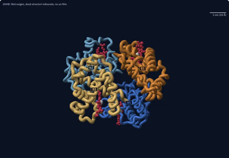
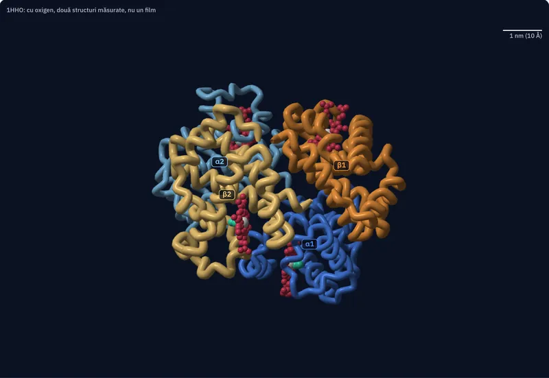
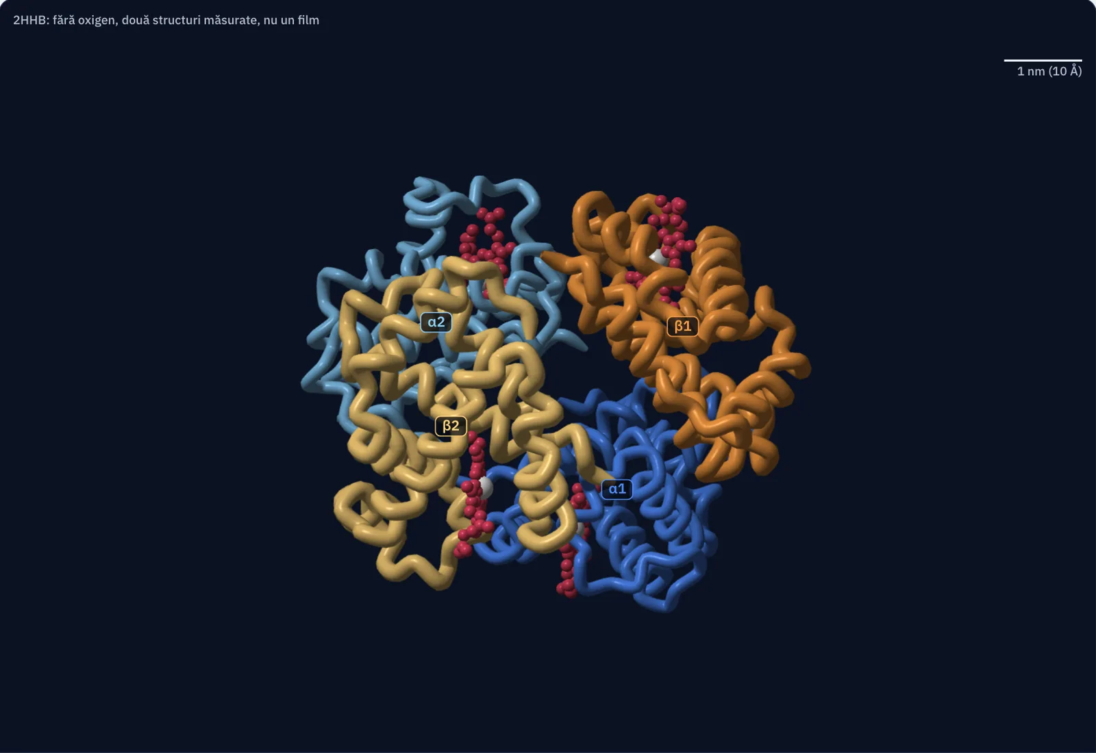
Haemoglobin in the red blood cells carries oxygen through the body. The picture beside the text is built from the positions of its atoms, measured with X-rays, in two states: without oxygen and with oxygen. Press “Add oxygen” and watch what changes.
Haemoglobin is measured atom by atom
Every chain in the picture is built from positions measured on a crystal of human haemoglobin: 4,384 protein atoms and 172 haem atoms, in structure 2HHB, at a resolution of 1.74 Å (1 Å is 0.1 nm).
Before you look closer: how many oxygen molecules can a single haemoglobin molecule carry?
A chain of 141 amino acids folds into a shape
A protein is a chain of amino acids. The chain does not lie stretched out: it packs into a compact shape, and the shape decides what the protein can do.
In the file, the alpha chain has 141 amino acids and 1,069 atoms, and the beta chain has 146 amino acids and 1,123 atoms. The tube follows the backbone of the chain, amino acid by amino acid.
Four chains, four haems, four iron atoms
The molecule has two alpha chains and two beta chains, 574 amino acids in all. Each chain holds a haem, a flat ring, red in the picture, with a white iron atom in the centre. Each iron atom has room for one O₂ molecule.
From the coordinates, the molecule without oxygen measures about 6.6 nm along its longest direction, between atom centres. A red blood cell is 7–8 µm across: between 1,100 and 1,200 molecules like this one would fit side by side across its diameter.
With oxygen, the iron moves almost into the plane of the haem
The alpha haem of chain α1, seen from the side. The disc is the plane of the four nitrogen atoms in the ring; the dots connect the iron to the plane. Without oxygen, the iron sits 0.45 Å and 0.34 Å from the plane in the two alpha haems (mean 0.40 Å), and 0.33 Å and 0.38 Å in the two beta haems (mean 0.36 Å), on the side of the histidine that links it to the chain.
With oxygen, in chains α1 and β1 the distances are 0.12 Å and −0.11 Å (the minus sign means on the side of the oxygen): the iron is almost in the plane. For scale, each nitrogen atom around the iron lies 2.0 Å from it.
The shift is small, at the limit of the precision of structures resolved to 1.74 and 2.1 Å, but it has the same direction in all the haems: with oxygen, the iron is closer to the plane. The means match the values published with the two structures (Fermi et al., 1984; Shaanan, 1983).
The pairs of chains rotate relative to each other
The iron is bonded to a histidine; when oxygen moves it, the histidine pulls the chain it belongs to, and the change is passed through the molecule.
The two structures were superposed on the α1β1 pair (287 amino acids, mean deviation 0.9 Å). The α2β2 pair fits over the other after a rotation of 13.0°. The picture looks along the axis of the rotation; the grey outline is the other state.
The distance between the irons of the two beta chains is 39.5 Å without oxygen and 34.6 Å with oxygen. Between the irons of the two alpha chains almost nothing changes: 34.2 Å and 34.7 Å. The values are calculated from the coordinates.
One change in the beta chain changes how the molecules bind to each other
At position 6 of each beta chain, the file has glutamic acid (Glu); its atoms are pink, on the surface of the molecule. In sickle cell anaemia, valine (Val) sits there instead, because a single base in the DNA is changed (MedlinePlus Genetics).
One amino acid out of the 146 in the beta chain changes how haemoglobin molecules bind to each other, and the red blood cells can take the shape of a sickle. The structures in the picture are of ordinary haemoglobin; sickle haemoglobin is not drawn here.
Now you explain
The picture shows the haem in chain β1, with O₂ bound to the iron; each of the four iron atoms has such a place. Carbon monoxide (CO), from combustion gases, binds to the same iron atom as oxygen, more strongly than oxygen does, and comes off very slowly (StatPearls, Physiology, Oxygen Transport; the article PMC11137813).
A person breathes air containing CO. Explain why their blood carries less oxygen, using what you saw about the binding sites on the iron atoms.
Compare with an answer
CO occupies the place at the iron atom where O₂ would be bound. A molecule has four such places, and every place taken by CO can no longer bind oxygen, so the blood carries less oxygen. CO comes off very slowly and also changes how haemoglobin releases oxygen to the tissues: the oxygen that is still bound is given up less readily.
The lesson on proteins, nucleic acids and ATP · The roles of proteins, for the Natural Sciences specialism