Chapter 1 · Investigating life
How do you find out how big something is when you cannot see it with the naked eye?
- Size
- from 1 mm to 1 nm
- Level
- cell and cell structures
- Time
- one lesson in the lab
Five real micrographs: two made with a light microscope, like the one in the school laboratory, and three with an electron microscope. Each has its own scale, and the scale tells you how big the thing you see is. Tap the image in two places: the length appears between the two ends.
The green structure on the right is a chloroplast
The chloroplast is the organelle where photosynthesis happens. This one sits in a cell of a bean leaf and was photographed with an electron microscope, in a very thin slice of the leaf.
Across the width of a pinhead, about 2 mm, 250 red blood cells fit in a line.
How many chloroplasts like this one, end to end, would fit across the same pinhead?
The scale bar turns pixels into micrometres
This is the skin of a red onion scale under a light microscope, magnified 100 times. The bar in the corner is labelled “200 µm” and measures 357 px in the original image.
The cell in the middle measures 227 px from one corner to the other. Its length: 227 ÷ 357 × 200 µm = 127 µm, which is 0.127 mm, because 1 mm = 1,000 µm.
The naked eye can tell two points apart if they are about 0.1 mm from each other, that is 100 µm. A red blood cell, at 8 µm, is 12.5 times smaller than this distance.
The membranes in the grana lie far below the limit of the light microscope
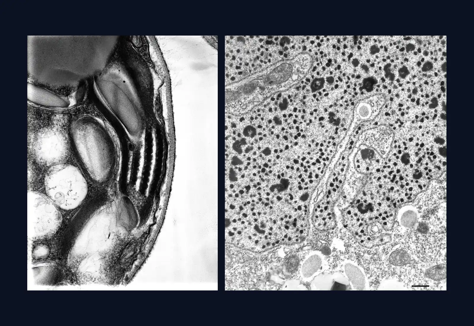
The chloroplast image is 4.5 µm wide, less than a single red blood cell. The chloroplast measures 3.41 µm from tip to tip.
The stacks of dark lines inside it are the grana, membranes laid one on top of another. The granum at bottom right is about 180 nm thick, and its membranes lie about 18 nm apart.
A school light microscope can separate points that are about 0.2 µm = 200 nm apart. The thickness of the granum is close to this limit, so under that microscope a granum appears at best as a green dot. The distance between the membranes is more than 11 times smaller: their structure can be resolved only with an electron microscope.
Digital zoom adds no detail
The segment on the eyespot of the alga Chlamydomonas is 1,000 pixels long in the original image. The source gives the size of one pixel: 0.8 nm. The length is 1,000 × 0.8 nm = 800 nm = 0.8 µm.
Enlarge the image eight times. On screen the segment becomes eight times longer, and the calculated length stays 0.8 µm, because the pixels of the original are the same.
Beyond a certain zoom, each pixel of the file covers several screen pixels and little squares appear. Digital zoom makes the image bigger without adding detail that the microscope did not record. The same happens when you enlarge a photo on a phone.
A section shows only a slice of the cell
For the electron microscope the cell is cut into very thin slices. The slice through Chlamydomonas is 250 nm thick, and the one through Paramecium about 75 nm.
The image shows a small part of the macronucleus of a Paramecium; the large dark spots are nucleoli. The whole macronucleus can be 35 µm long and 12 µm wide, so the edges of the image cut through it.
Measuring is switched off here. The author printed the negative on paper and then scanned it, and the source says the image is suitable for qualitative analysis. The 0.5 µm bar in the corner gives only the order of magnitude.
The green of the bean chloroplast was added by people
Electrons have no colour, so an electron micrograph comes out in shades of grey, like the Chlamydomonas image. The right-hand half shows the same bean image turned grey.
In a light microscope the colours belong to the specimen: the red of the onion cells comes from their pigment, the green of Spirogyra from chlorophyll.
Who coloured the bean chloroplast green?
Now you explain
A classmate photographs the onion cells in the school microscope with a phone, at 400×, then enlarges the photo eight times on screen and says: “Now I can see the ribosomes too.”
Ribosomes are 10–30 nm. What do you answer?
Compare with an answer
A school light microscope separates two points only if they are about 0.2 µm = 200 nm apart or further. Ribosomes, at 10–30 nm, are far below this limit, so their structure does not appear in the image the phone captured. Digital zoom stretches the pixels of that image: the photo gets bigger, but it gets no new detail. The structure of ribosomes can be resolved with an electron microscope.
The lesson on the light microscope, with the worked example and questions
