Chapter 3 · The cellLesson 3.4
Cell organelles: ribosomes, reticulum, Golgi, lysosomes, mitochondria, plastids, vacuoles and centrosome
Why does a green tomato turn red as it ripens, and what has actually changed in its cells?
- Size
- from 5 nm to 100 µm
- Level
- cell
- Time
- a day in the life of a cell
Before you read: what do you think?
A cell in a plant leaf has chloroplasts, which it uses for photosynthesis. Does it also have mitochondria?
See the answer
Yes. Chloroplasts use light to make sugars, and mitochondria break sugars down and produce ATP, day and night. Plants need both, and mitochondria are also found in root cells, where light does not reach.
The flesh cells of a green tomato contain chloroplasts with chlorophyll. As it ripens, the chlorophyll is broken down and the chloroplasts turn into chromoplasts, which collect red and orange pigments; in a tomato, lycopene even builds up as crystals. The colour of the fruit shows that the organelles in its cells have changed jobs.
The organelles that make proteins and send them where they belong
Ribosomes are small organelles without a membrane, made of RNA and proteins, in two subunits. On them, amino acids are linked into chains: this is where proteins are made. Some float free in the cytosol and make proteins used right there; others are attached to the membrane of the endoplasmic reticulum.
The endoplasmic reticulum (ER) is a network of flattened sacs and channels enclosed by membrane. The rough ER has ribosomes on the side facing the cytosol: the proteins they make enter the reticulum, where they fold and are modified. The rough ER also makes phospholipids for membranes. The smooth ER has no ribosomes; it makes lipids and steroid hormones, neutralises some medicines and poisons, and in muscle cells it stores calcium ions.
The Golgi apparatus is a stack of flattened sacs. It receives proteins and lipids from the reticulum in small vesicles, modifies them, sorts them and packs them into other vesicles. Some of these fuse with the plasma membrane and empty their contents outside the cell; in other words, they secrete them.
Lysosomes are vesicles with enzymes that break down proteins, carbohydrates, lipids and nucleic acids. They digest worn-out parts of the cell and bacteria swallowed by defence cells. They are typical of animal cells; in a plant cell, the vacuole does this job.
The energy organelles: the mitochondrion and the plastids
The mitochondrion has two membranes. The inner one forms folds called cristae, which surround a ground substance called the matrix. Most of cellular respiration takes place in the mitochondrion: the energy in sugars is transferred to ATP, the molecule the cell uses as its energy source. Mitochondria are found in almost all eukaryotic cells, including those of plants and fungi, and have their own DNA and ribosomes.
Plastids are double-membrane organelles found in plants and algae. Chloroplasts contain chlorophyll and carry out photosynthesis: inside them are flattened membrane sacs, the thylakoids, arranged in stacks called grana and surrounded by the stroma. Chromoplasts collect carotenoid pigments, yellow, orange and red, and give many flowers and fruits their colour. Leucoplasts are colourless and store substances; among them, amyloplasts store starch in roots, seeds and tubers such as the potato.
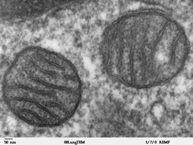
Photo: Louisa Howard, Dartmouth Electron Microscope Facility, Public domain, source. Unchanged.

| Slice | Height in the volume | Nucleus area | Mitochondrial profiles |
|---|---|---|---|
| 1 of 60 | 0.5 µm | does not appear | 10 |
| 7 of 60 | 1.1 µm | 69 µm² | 107 |
| 13 of 60 | 1.6 µm | 182 µm² | 110 |
| 19 of 60 | 2.1 µm | 220 µm² | 88 |
| 24 of 60 (widest nucleus) | 2.6 µm | 225 µm² | 73 |
| 31 of 60 | 3.2 µm | 217 µm² | 21 |
| 37 of 60 | 3.8 µm | 191 µm² | 11 |
| 43 of 60 | 4.4 µm | 143 µm² | 5 |
| 49 of 60 | 4.9 µm | 78 µm² | 7 |
| 60 of 60 | 5.9 µm | does not appear | 0 |
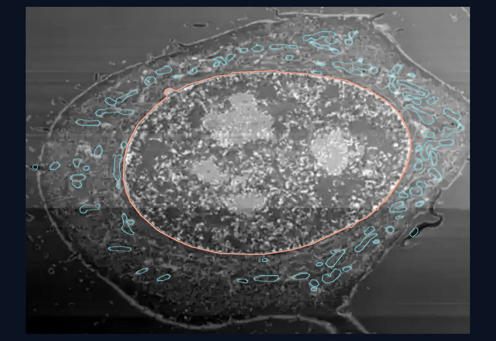
The nucleus appears in 53 of the 60 slices; it measures 21.2 µm × 13.6 µm and is 5.0 µm thick, in a volume 6.4 µm tall. In slice 24, the program found 73 mitochondrial profiles. The frame is 32.8 µm × 24.1 µm; the distance between slices is 91 nm.
Micrograph and calculation. Micrograph: 60 planes through the volume of a HeLa cell, measured with the FIB-SEM electron microscope (dataset jrc_hela-2, OpenOrganelle, Janelia Research Campus, CC BY 4.0). Each plane is calculated from the original images, which were taken in a different direction, layer by layer; grey means electron density, not colour. The brightness is adjusted separately in each plane, and 3–5 rows of pixels in each plane, brighter than their neighbours, were replaced with the mean of the neighbours. Zoom adds no detail: one displayed pixel is 64 nm. The outlines are predictions made by a program (automatic segmentation) and can be wrong. The bacterium is a drawing to scale, not a micrograph.
Sources and credits. Janelia Research Campus, the COSEM project, dataset jrc_hela-2 (FIB-SEM electron microscopy and automatic segmentations), published through OpenOrganelle and through Cell Organelle Segmentation in Electron Microscopy (COSEM) on AWS, licence CC BY 4.0 (source), registered at source and at source; segmentation method: Heinrich et al., Whole-cell organelle segmentation in volume electron microscopy, Nature, 2021. The images are reduced in size, with the brightness adjusted, and cut in a different plane. Chloroplast in a bean leaf, TEM micrograph, artificially coloured: Kevin Mackenzie, University of Aberdeen, Wellcome Collection B0009849, CC BY 4.0, reduced in size. Size of the bacterium: Escherichia coli, about 2.0 µm long and 0.25–1.0 µm in diameter (drawn at 0.5 µm), from Wikipedia, the article Escherichia coli.
Vacuoles and the centrosome
Vacuoles are membrane-bound sacs that store water and substances. A mature plant cell usually has a large central vacuole, which takes up most of its volume, regulates the amount of water and keeps the cell firm. Animal cells have a few small vacuoles.
The centrosome lies next to the nucleus, in animal cells. It is made of two centrioles set at right angles to each other, each a cylinder of nine groups of three microtubules. The centrosome organises the cell’s microtubules and, during division, the spindle that separates the chromosomes. Most plant cells have no centrioles and organise their microtubules in another way.
Plant, animal and fungal cells compared
All three are eukaryotic: they have a nucleus, ribosomes, endoplasmic reticulum, a Golgi apparatus and mitochondria. The differences asked about in tests are few and clear-cut.
A plant cell has a cellulose cell wall, plastids (chloroplasts, chromoplasts, leucoplasts) and a large central vacuole, and stores carbohydrates as starch. Most plant cells have no centrioles.
An animal cell has no wall and no plastids, has a centrosome with centrioles, lysosomes and small vacuoles, and stores carbohydrates as glycogen.
A fungal cell has a cell wall made of chitin, has no chloroplasts and no chlorophyll, has vacuoles and, like animals, stores glycogen. In its membrane, the place of the cholesterol found in animals is taken by a similar substance, ergosterol.
Worked example
The route a protein takes and the time between stages
In 1964, Lucien Caro and George Emil Palade, the Romanian-born cell biologist, injected guinea pigs with a radioactive amino acid. Cells in the pancreas built it into the proteins they were making at that moment, and the researchers then looked under the microscope for the labelled proteins after different intervals.
The result, in the living guinea pig: the labelled protein was in the reticulum after about 5 minutes, in the Golgi apparatus after about 20 minutes and in the secretory granules after an hour.
Step 1: between the observation in the reticulum and the one in the Golgi apparatus, about 20 − 5 = 15 minutes passed.
Step 2: between the observation in the Golgi apparatus and the one in the granules, about 60 − 20 = 40 minutes passed.
These are intervals between observations; the journey of a single protein was not timed.
Step 3: interpret. The protein is not made in the Golgi apparatus, nor does it appear directly in the granules. It passes, in order, through the ribosomes on the reticulum, through the reticulum and through the Golgi apparatus, and reaches the granules, from which it is released outside the cell.
In Romania
George Emil Palade and the grains on the membranes
George Emil Palade was born in Iași, in north-eastern Romania, on 19 November 1912. Using the electron microscope, he showed in 1953 that nearly all the cells he examined have small grains, 10–15 nm across, many of them attached to the membranes of the endoplasmic reticulum. The paper came out in 1955. Others had seen similar particles before; his contribution was to describe them systematically, in more than 40 types of cell.
The role of the grains was proved later: in 1960, Palade and his colleagues showed, along with other groups, that they link amino acids into proteins. The name “ribosome” was suggested at a symposium in 1958, and the sources do not say who proposed it; for a short time, ribosomes were also called “Palade granules”.
In 1974, the Nobel Prize in Physiology or Medicine was shared in three equal parts between Albert Claude, Christian de Duve, who had described lysosomes, and George E. Palade, “for their discoveries concerning the structural and functional organization of the cell”.
In short
- Ribosomes, which have no membrane, make proteins; the rough ER, with ribosomes, receives and modifies proteins, and the smooth ER makes lipids and neutralises some substances.
- The Golgi apparatus modifies, sorts and packs proteins into vesicles, some of which are sent outside the cell; lysosomes digest worn-out parts and swallowed bacteria.
- The mitochondrion has two membranes, the inner one with cristae, and produces most of the ATP through cellular respiration, in plants, animals and fungi.
- The plastids are chloroplasts (photosynthesis), chromoplasts (carotenoid pigments) and leucoplasts (storage, of starch for example).
- A plant cell has a large central vacuole, and in an animal cell the centrosome with its two centrioles organises the microtubules.
- A plant cell has a cellulose wall and plastids, an animal cell has no wall and has centrioles, and a fungal cell has a wall made of chitin and no chloroplasts.
Common mistakes
WrongPlants have chloroplasts instead of mitochondria.
RightPlants have both chloroplasts and mitochondria. Chloroplasts use light to make sugars, and mitochondria break them down and produce ATP.
WrongProteins are made in the Golgi apparatus.
RightProteins are made on ribosomes. The Golgi apparatus receives them from the reticulum, modifies them, sorts them and sends them on.
WrongAll the cells of a plant have chloroplasts.
RightOnly the cells in green parts have chloroplasts. Cells in the root or in a potato tuber have leucoplasts, and those in petals and ripe fruit often have chromoplasts.
Check yourself
Seven questions. For the first six you see the explanation at once, with the paragraph of the lesson that supports it; for the written ones you compare with a good answer.
Which organelle assembles the amino acid chain of a protein?
Answer
b) The ribosome. Amino acids are linked into proteins on ribosomes. The Golgi apparatus only modifies and sorts proteins that are already made.
The cells of a potato tuber are full of starch, though they are not green. What plastids do they have?
Answer
d) Leucoplasts (amyloplasts). Leucoplasts are colourless storage plastids, and amyloplasts store starch. Mitochondria are not plastids.
Which organelle is missing from most plant cells but found in the animal cell?
Answer
a) The centrosome with centrioles. Animal cells have a centrosome with two centrioles; most plant cells have no centrioles. Mitochondria, the Golgi apparatus and the reticulum are found in both.
In the 1964 experiment, the labelled protein was in the Golgi apparatus after about 20 minutes and in the granules after an hour. About how many minutes passed between the two observations?
minutesAnswer
40 minutes. An hour is 60 minutes: 60 − 20 = 40 minutes.
Put in order the places a protein passes through on its way out of the cell.
Answer
The correct order:
- The ribosomes on the rough endoplasmic reticulum
- The inside of the endoplasmic reticulum
- Small vesicles heading for the Golgi apparatus
- The Golgi apparatus
- Secretory granules
- Outside the cell
The protein is made on the ribosomes attached to the reticulum, enters the reticulum, reaches the Golgi apparatus in vesicles and is packed into granules, which empty outside the cell.
Choose the type of cell each feature belongs to.
Answer
Plant cell: Cellulose cell wall; Chloroplasts and a large central vacuole; Starch as the storage substance.
Animal cell: No cell wall; Centrosome with two centrioles.
Fungal cell: Cell wall made of chitin; Ergosterol in the membrane.
The cellulose wall, chloroplasts and starch belong to the plant cell; the lack of a wall and the centrioles, to the animal cell; the chitin wall and ergosterol, to the fungal cell.
Cells in the pancreas send out large amounts of digestive enzymes, which are proteins. Which organelles do you expect to be highly developed in them, and why?
A good answer
I expect a lot of rough endoplasmic reticulum, with many ribosomes, because that is where export proteins are made and modified. The Golgi apparatus must be large, to sort the enzymes and pack them into secretory granules. And many mitochondria, because making and transporting proteins uses ATP.
A complete answer links the function (protein secretion) to the rough ER with ribosomes and to the Golgi apparatus, explains the role of each and may add mitochondria as the source of ATP.
Terms
- organelle
- A compartment of the cell with a precise structure and role, usually enclosed by a membrane.
- endoplasmic reticulum
- A network of membrane-bound sacs and channels; the rough ER has ribosomes and processes proteins, the smooth ER makes lipids.
- Golgi apparatus
- A stack of flattened sacs that modifies, sorts and packs proteins and lipids into vesicles.
- lysosome
- A vesicle with digestive enzymes that breaks down worn-out parts of the cell and swallowed material.
- mitochondrion
- An organelle with two membranes, the inner one with cristae, in which most of the ATP is produced through cellular respiration.
- plastids
- Double-membrane organelles of plants and algae: chloroplasts, chromoplasts and leucoplasts.
- vacuole
- A membrane-bound sac that stores water and substances; in a mature plant cell it is usually large and central.
- centrosome
- An organelle of the animal cell, made of two centrioles, that organises the microtubules and the division spindle.
Sources and link to the programme
- OpenStax – Biology 2e, 4.3 Eukaryotic Cells – free and ER-bound ribosomes make proteins; the mitochondrion with two membranes, cristae, matrix, ATP, its own DNA and ribosomes; chloroplasts with thylakoids, grana, stroma, chlorophyll; the central vacuole takes up most of the volume; the centrosome with two perpendicular centrioles, made of nine triplets of microtubules; lysosomes in animals, digestion in the vacuole in plants; the differences between the animal cell and the plant cell
- OpenStax – Biology 2e, 4.4 The Endomembrane System and Proteins – rough ER (ribosomes, folding and modification of proteins, phospholipids); smooth ER (lipids, steroid hormones, detoxification, calcium storage in muscle); the Golgi apparatus (sorting, packaging, secretion); lysosomes (hydrolytic enzymes, digestion of engulfed pathogens)
- OpenStax – Biology 2e, 24.1 Characteristics of Fungi – chitin wall, glycogen storage, no chloroplasts and no chlorophyll, ergosterol instead of cholesterol
- OpenStax – Biology 2e, 3.2 Carbohydrates – starch is the storage form of carbohydrates in plants, glycogen in animals
- Sadali, Sowden, Ling, Jarvis – Differentiation of chromoplasts and other plastids in plants, Plant Cell Reports 2019 (PMC) – chromoplasts accumulate carotenoids, in flowers and fruits; in tomato, lycopene crystals; chromoplasts can derive from chloroplasts, with the breakdown of chlorophyll, during ripening; colourless leucoplasts; amyloplasts store starch in roots, seeds and tubers
- Karolinska Institutet – Press release: The Nobel Prize in Physiology or Medicine 1974 – the prize shared between Claude, de Duve and Palade; lysosomes identified by de Duve; Palade showed, along with other groups, the role of the particles in protein synthesis
- NobelPrize.org – George E. Palade, Facts – the prize motivation, “for their discoveries concerning the structural and functional organization of the cell”; the division into three equal parts
- George E. Palade – Autobiografie, Les Prix Nobel 1974 – born in Iași, 19 November 1912
- G. E. Palade – A small particulate component of the cytoplasm, J Biophys Biochem Cytol 1(1):59–68, 1955 (PMC) – the 1953 presentation and the 1955 paper; grains of 100–150 Å (10–15 nm); more than 40 cell types; similar particles seen and isolated earlier by others
- Caro, Palade – Protein synthesis, storage, and discharge in the pancreatic exocrine cell, J Cell Biol 1964 (PMC) – radioactive leucine in the living guinea pig; the labelled protein in the reticulum after about 5 minutes, in the Golgi after about 20 minutes, in the granules after an hour
- R. B. Roberts (ed.) – Microsomal Particles and Protein Synthesis, 1958 (Internet Archive) – the word “ribosome” was suggested during the February 1958 symposium, without saying by whom
- Marilyn G. Farquhar – George E. Palade 1912–2008, Biographical Memoirs, National Academy of Sciences – the name “Palade granules”, used for a short time for ribosomes
From the programme: “Fundamental cell types - prokaryotes and eukaryotes. Components of the cell – the main structural elements and their functions: membrane/cell wall, cytoplasm, cell organelles (ribosomes, mitochondria, Golgi apparatus, ER, lysosomes, plastids, vacuoles, centrosome), nucleus/nucleoid” (common core). Specific competences addressed: 1.1, 2.1, 2.2.