Chapter 3 · The cellLesson 3.3
Membrane, cell wall, cytoplasm and nucleus: the cell’s coverings and its centre
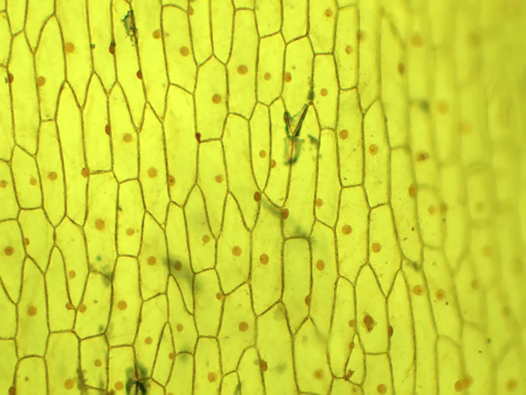
Under the school microscope, onion skin shows the outlines of cells and a darker dot in each one. What are they, and why can’t you see the membrane?
- Size
- from 5 nm to 100 µm
- Level
- cell
- Time
- a day in the life of a cell
Before you read: what do you think?
The plasma membrane is often called the cell’s “lipid layer”. How much of the mass of the membrane of an ordinary human cell is protein?
See the answer
About half. Lipids form the framework of the membrane, a double layer, but the proteins held in it weigh roughly as much as the lipids and carbohydrates together. Proteins do much of the membrane’s work: they transport substances, receive signals and link cells to one another.
Put a scrap of the thin skin from the inside of an onion layer on a slide and look at it under the microscope: you see something like a brick wall, with a darker dot in each brick. The outlines you see are the cell walls, and the dots are the nuclei. The membrane of each cell is there, pressed against the wall, but it is too thin to see.
The plasma membrane: a double layer of lipids with proteins held in it
The plasma membrane (plasmalemma) surrounds every cell and is 5–10 nm thick, tens of times below what a light microscope can separate. Its framework is a double layer of phospholipids. Each phospholipid has a “head” that attracts water and two “tails” that repel it. In the double layer, the heads face the water outside and the cytoplasm, and the tails face each other in the middle.
Proteins are held in the lipid layer. Some cross it completely and are called integral proteins; others sit on its surface and are called peripheral proteins. In an ordinary human cell, proteins make up about half the mass of the membrane. The membrane of animal cells also contains cholesterol, and chains of sugars attached to its outer face form the glycocalyx.
In 1972, S. J. Singer and Garth Nicolson described the membrane with the fluid mosaic model. “Mosaic”, because it is made of different molecules set side by side. “Fluid”, because the lipids and many proteins can move within the plane of the membrane. The membrane is partially permeable (selectively permeable): it lets some substances through easily, and stops others or lets them through only by way of its transport proteins.
The cell wall: outside the membrane, not instead of it
The cells of plants, of fungi and of most bacteria have a rigid cell wall over the membrane, which gives the cell its shape, supports it and protects it. The substance it is made of differs: in plants, cellulose; in fungi, chitin, the same substance found in the shells of insects and crayfish; in bacteria, murein. Animal cells have no wall.
The wall does not replace the membrane. A plant cell has both a wall and a membrane, and the membrane, pressed against the wall, controls what enters the cell. The wood of a tree is made largely of the walls of dead cells, thickened and hardened with a substance called lignin.

The cytoplasm: cytosol, cytoskeleton and organelles
The cytoplasm is everything between the plasma membrane and the nucleus. It has three parts. The cytosol (hyaloplasm) is the jelly-like part, made mostly of water with dissolved ions, sugars, amino acids and enzymes; many of the reactions of metabolism take place in it. The cytosol holds the organelles, compartments with a precise structure and role, and in a eukaryotic cell all of this is supported by an internal skeleton.
The cytoskeleton is a network of protein fibres. Microfilaments, made of actin, are about 7 nm thick and take part in cell movement, in changes of cell shape and in the currents of the cytoplasm. Intermediate filaments, 8–10 nm thick, hold the cell’s shape and anchor the nucleus. Microtubules, about 25 nm thick, are tracks along which vesicles travel, and they pull the chromosomes apart during division; cilia and flagella are made of them too.
The nucleus: where a eukaryotic cell keeps its DNA
The nucleus is surrounded by a nuclear envelope made of two membranes. The envelope has nuclear pores, through which ions, molecules and RNA pass between the nucleus and the cytoplasm. The inside of the nucleus is called the nucleoplasm.
The DNA in the nucleus is bound to proteins and forms chromatin, which in a cell that is not dividing looks like a tangle of thin threads. Before division, the chromatin condenses into chromosomes. After staining, a darker area shows up in the nucleus, the nucleolus, where ribosomal RNA joins with proteins and the ribosome subunits are formed.
Through the information in DNA, the nucleus controls which proteins the cell makes, and therefore what the cell does. Prokaryotes have no nucleus: their DNA lies in the nucleoid, with no envelope.
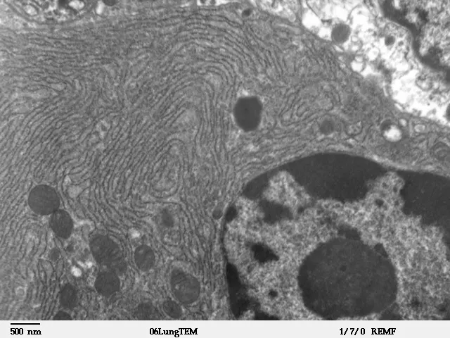
Photo: Louisa Howard, Dartmouth Electron Microscope Facility, Public domain, source. Unchanged.

Worked example
The onion skin slide
With tweezers, peel the thin skin from the inner face of an onion layer and spread it on a slide, in a drop of water. Add a drop of stain, for example iodine solution (Lugol’s iodine) or methylene blue, cover with a coverslip and look first with the low-power objective, then with the highest-power one.
What you see: elongated cells, pressed against one another. The thick lines between them are the cell walls, made of cellulose. In each cell, the nucleus appears as a round, darker-stained body, sometimes with one or two nucleoli. The cytoplasm forms a thin layer next to the wall, and the centre of the cell is taken up by a large vacuole.
What you do not see: the plasma membrane. It is at most 10 nm thick, and the light microscope cannot separate details smaller than about 200 nm, so the membrane is at least 20 times below its limit. It is there, pressed against the wall, and you can make it visible indirectly: in a very salty solution, the contents of the cell come away from the wall, and their edge shows where the membrane is.
In Romania
Maya and Nicolae Simionescu and the membrane of blood vessels
Between 1970 and 1979, the Romanian researchers Nicolae and Maya Simionescu worked with George Emil Palade, the Romanian-born cell biologist, at Rockefeller University and at Yale University. They studied the membrane of the cells that line the inside of capillaries. These cells are very thin and full of small vesicles pinched off from the plasma membrane.
Using marker molecules of known sizes, some of them 4.5–30 nm, they showed together with Palade that the vesicles carry substances from one side of the cell to the other, from the blood towards the tissue. This kind of transport is called transcytosis. They also showed that the side of the membrane facing the blood is not the same everywhere: it has small areas with a different chemical make-up.
In 1979, the two opened the Institute of Cellular Biology and Pathology in Bucharest; Palade, Christian de Duve and Günter Blobel came to its inauguration. Today the institute bears Nicolae Simionescu’s name.
In short
- The plasma membrane is 5–10 nm thick and is a double layer of phospholipids holding integral and peripheral proteins; the fluid mosaic model (Singer and Nicolson, 1972) describes it as a mosaic of molecules that can move.
- The cell wall lies outside the membrane and is made of cellulose in plants, chitin in fungi and murein in bacteria; animal cells have no wall.
- The cytoplasm comprises the cytosol (hyaloplasm), the organelles and the cytoskeleton, made of microfilaments, intermediate filaments and microtubules.
- The nucleus has a double envelope with pores and contains chromatin (DNA bound to proteins) and the nucleolus, where the ribosome subunits are formed.
- Prokaryotes have no nucleus: their DNA lies in the nucleoid, with no envelope.
Common mistakes
WrongA plant cell has a wall instead of a membrane.
RightA plant cell has both a wall and a membrane. The wall sits outside the membrane and supports it, and the membrane controls exchanges with the surroundings.
WrongThe cytoplasm is water in which organelles float at random.
RightThe cytosol is a gel with many dissolved substances, crossed by the cytoskeleton, which holds the organelles in place and moves them.
WrongThe cell membrane is made only of fats.
RightPhospholipids form the framework of the membrane, but in an ordinary human cell proteins make up about half its mass.
Check yourself
Seven questions. For the first six you see the explanation at once, with the paragraph of the lesson that supports it; for the written ones you compare with a good answer.
What is the cell wall of a mushroom made of?
Answer
c) Chitin. Fungi have walls made of chitin. Cellulose is in plant walls, murein in bacterial walls, and phospholipids form the membrane.
How are the phospholipids arranged in the plasma membrane?
Answer
b) In two layers, with the tails facing each other in the middle. The heads attract water and face the outside and the cytoplasm; the tails repel water and shelter in the middle of the double layer.
What is formed in the nucleolus?
Answer
a) The ribosome subunits. In the nucleolus, ribosomal RNA joins with proteins to form the ribosome subunits, which then leave through the nuclear pores.
A microtubule is about 25 nm thick and a microfilament about 7 nm. How many times thicker is the microtubule? Round to one decimal place.
timesAnswer
3.6 times. 25 ÷ 7 ≈ 3.6. Microtubules are the thickest fibres of the cytoskeleton, and microfilaments the thinnest.
Put the steps for the onion skin slide in order.
Answer
The correct order:
- Peel the skin from the inner face of the onion layer
- Spread it on the slide, in a drop of water
- Add a drop of stain
- Cover with the coverslip
- Look first with the low-power objective, then with the high-power one
The slide is first made wet and stained, then covered, and at the microscope you start with low magnification to find a good area.
Why can’t you see the plasma membrane under the school light microscope?
Answer
c) Because it is at most 10 nm thick, far below the light microscope’s limit of about 200 nm. Every cell has a membrane, but it is at least 20 times thinner than the smallest detail the light microscope can separate.
A medicine destroys only the cell wall. What would happen to a bacterial cell, and what would happen to a cell of the human body? Explain.
A good answer
The bacterium would lose the support outside its membrane; left with only the membrane, it could swell and burst, in water for example. The human cell would not be affected, because animal cells have no wall; its membrane stays whole. A medicine that targets the wall therefore hits the bacterium and spares human cells.
A complete answer says that bacteria have a wall (made of murein) and animal cells do not, that the wall supports the membrane without replacing it, and draws a conclusion about the different effect on the two cells.
Practical work for this lesson
- The micrograph: observation and scale – Observation of electron micrographs, 30–40 minutes
Terms
- plasma membrane
- The thin covering, 5–10 nm thick, that encloses every cell and controls its exchanges with the surroundings.
- fluid mosaic model
- The description of the membrane as a double layer of phospholipids holding proteins, whose molecules can move within the plane of the membrane.
- cell wall
- A rigid covering outside the membrane, made of cellulose in plants, chitin in fungi and murein in bacteria.
- cytosol
- The jelly-like part of the cytoplasm, also called hyaloplasm, which holds the organelles.
- cytoskeleton
- The network of protein fibres in the cytoplasm (microfilaments, intermediate filaments, microtubules) that holds the cell’s shape and moves the organelles.
- nuclear envelope
- The two membranes that enclose the nucleus, crossed by nuclear pores.
- chromatin
- The DNA of the nucleus bound to proteins, in the form of thin threads in a cell that is not dividing.
- nucleolus
- The area of the nucleus where the ribosome subunits are formed.
Sources and link to the programme
- OpenStax – Biology 2e, 5.1 Components and Structure – the fluid mosaic model, Singer and Nicolson, 1972; the 5–10 nm thickness; phospholipids with hydrophilic heads and hydrophobic tails; integral and peripheral proteins; cholesterol; glycocalyx; proteins make up about 50% of the membrane mass of a typical human cell
- OpenStax – Biology 2e, 4.3 Eukaryotic Cells – the cytoplasm between the membrane and the nuclear envelope; the jelly-like cytosol; double nuclear envelope with pores for ions, molecules and RNA; chromatin; the nucleolus and the ribosomal subunits; the cellulose cell wall in plants, also present in fungi
- OpenStax – Biology 2e, 4.5 The Cytoskeleton – microfilaments of about 7 nm (actin), intermediate filaments of 8–10 nm, microtubules of about 25 nm and their roles; cilia and flagella are made of microtubules
- OpenStax – Biology 2e, 24.1 Characteristics of Fungi – the fungal wall is made of chitin, the substance of the arthropod exoskeleton
- OpenStax – Microbiology, 3.3 Unique Characteristics of Prokaryotic Cells – the bacterial wall made of peptidoglycan (murein)
- OpenStax – Biology 2e, 30.2 Stems – tracheids and vessel elements are dead at maturity and have thickened, lignified walls
- OpenStax – Biology 2e, 4.1 Studying Cells – the resolution of the light microscope, about 200 nm
- Marilyn G. Farquhar – George E. Palade 1912–2008, Biographical Memoirs, National Academy of Sciences – Palade’s collaboration with Nicolae and Maya Simionescu at Rockefeller and Yale; endothelial vesicles and transcytosis; markers of 4.5–30 nm (dextrans, glycogen); microdomains of different chemical make-up on the endothelial membrane; the inaugural symposium in Bucharest in September 1979
- Institutul de Biologie și Patologie Celulară „Nicolae Simionescu” – History – the collaboration with Palade at Rockefeller and Yale, 1970–1979; the 1979 inauguration, with Palade, de Duve and Blobel; the institute was named after Nicolae Simionescu in 1995
From the programme: “Fundamental cell types - prokaryotes and eukaryotes. Components of the cell – the main structural elements and their functions: membrane/cell wall, cytoplasm, cell organelles (ribosomes, mitochondria, Golgi apparatus, ER, lysosomes, plastids, vacuoles, centrosome), nucleus/nucleoid” (common core). Specific competences addressed: 1.1, 2.1, 2.2.