Chapter 2 · Molecules of lifeLesson 2.3
Proteins, nucleic acids and ATP: building, information, energy
How can a cell build thousands of different proteins from only 20 amino acids, and how does it know in what order to join them?
- Size
- from 0.1 nm to 10 nm
- Level
- atoms and molecules
- Time
- an instant
Before you read: what do you think?
Your cells use ATP non-stop. How much ATP does your body make and use in a single day?
See the answer
About as much as your whole body mass. The same molecules are rebuilt and used again many times a day: ATP splits into ADP and phosphate when it releases energy, then is rebuilt from them, mainly with energy from cellular respiration.
Hair, nails, the enzymes in saliva, the haemoglobin in blood and the antibodies that protect you from microbes are all proteins. They are built from the same 20 pieces, joined in different orders. The order is written in DNA.
Proteins are chains of amino acids
Proteins are organic substances made of units called amino acids. The proteins of living things normally contain 20 amino acids. The human body can make 11 of them; the other nine are called essential amino acids and must come from food.
Two amino acids join by a peptide bond; a molecule of water is removed as it forms. A chain of many amino acids is called a polypeptide. The order of amino acids in the chain is the protein’s primary structure. The chain then folds into a three-dimensional shape, and the protein’s role depends on that shape.
Heat, acids or other substances can spoil a protein’s shape without breaking the chain of amino acids: the protein is denatured and can no longer do its job. Egg white going from clear to white in a frying pan is an example of denaturation that cannot be reversed.
For the curious
In sickle cell anaemia, a single amino acid in the beta chain of haemoglobin is replaced by another, because of a change in a single base in DNA. The altered haemoglobin molecules can link up into long fibres, especially when the blood holds little oxygen; the fibres distort the red blood cells, which take on a sickle shape and can block small vessels.
What proteins do
Proteins have more roles than any other molecules in the cell. They have a structural role: keratin in hair and nails, collagen in skin and tendons, actin in muscle. They act as enzymes: they catalyse, that is, speed up, the chemical reactions in the cell. They have a transport role: haemoglobin carries oxygen. They have a defence role: antibodies recognise and bind microbes. Some proteins are hormones; insulin has two chains, one of 21 and one of 30 amino acids.
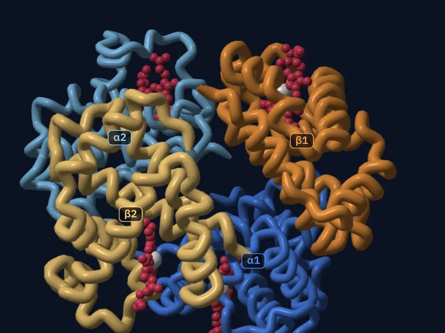
Haemoglobin shows clearly how shape and role are linked. A haemoglobin molecule is made of four chains of amino acids, two alpha chains and two beta chains. Each chain holds a haem group, a ring with an iron atom at its centre. Each iron atom binds one oxygen molecule, so one haemoglobin molecule can carry four molecules of O₂. This is where the iron in food, one of the mineral salts, ends up.
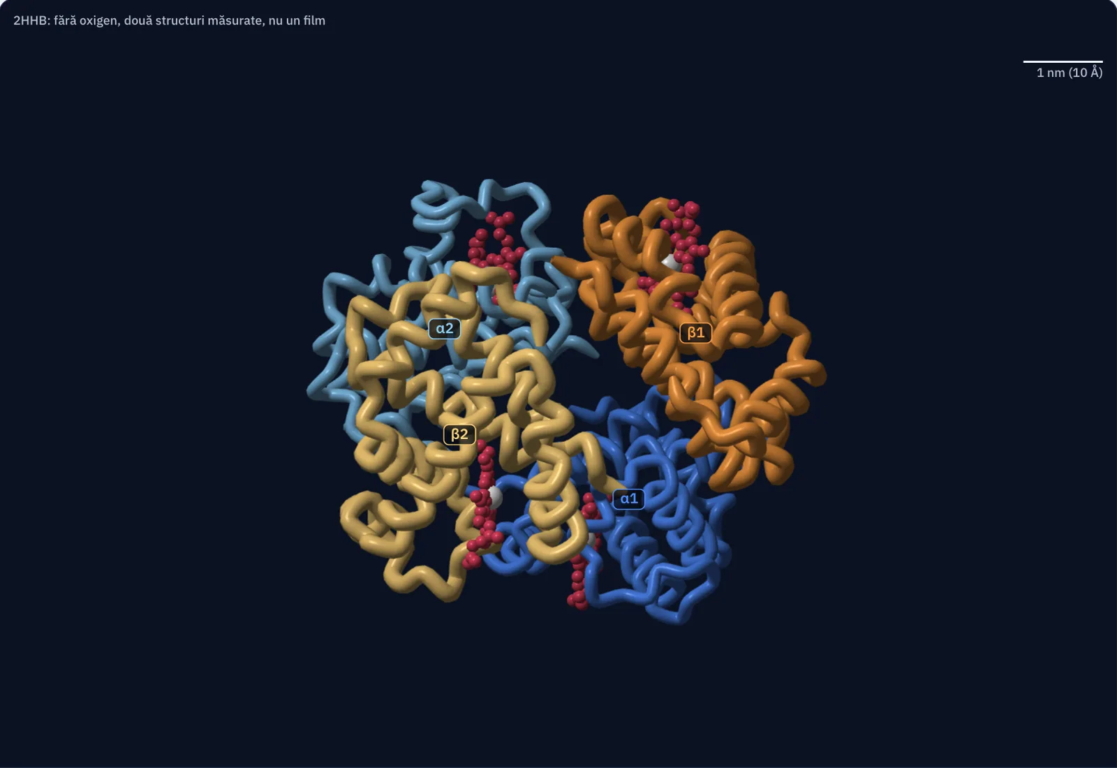
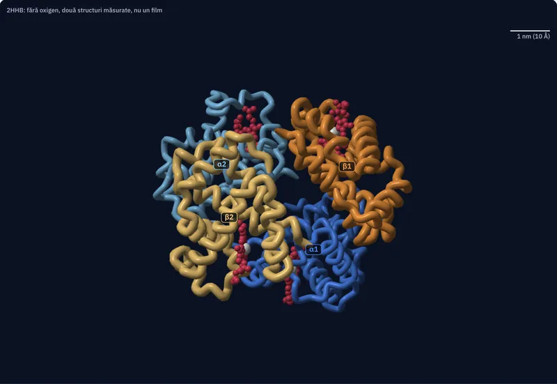
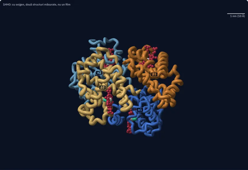
| What is measured, or calculated from the coordinates | Without oxygen (2HHB) | With oxygen (1HHO) |
|---|---|---|
| Amino acids: alpha chain, beta chain, total | 141, 146, 574 | 141, 146, 574 |
| Atoms of protein, of haem, of O₂ | 4,384, 172, 0 | 4,384, 172, 8 |
| Iron relative to the plane of the four nitrogen atoms: alpha haem, beta haem (Å) | 0.40, 0.36 | 0.12, −0.11 |
| Distance between the irons of the two beta chains (Å) | 39.5 | 34.6 |
| Distance between the irons of the two alpha chains (Å) | 34.2 | 34.7 |
| Longest extent of the molecule, between atom centres (nm) | 6.6 | 6.5 |
| Rotation of the α2β2 pair between the two structures, with α1β1 superposed | 13.0° | the same rotation |
Measurements and calculation. Measurement: the positions of the atoms, from structures 2HHB (without oxygen, 1.74 Å) and 1HHO (with oxygen, 2.1 Å), obtained by X-ray diffraction on crystals of human haemoglobin (RCSB Protein Data Bank, public domain). The two images are two structures, not a recording of a movement. In 1HHO the file contains a single alpha chain and a single beta chain; the second pair is a copy generated by crystal symmetry, as shown in the biological assembly. Calculation: the superposition of the two structures on the α1β1 pair, the distances and the angles. Drawing: the tubes, the spheres and the colours, chosen by the person who draws; the spheres have the radii of the atoms, and the iron and the O₂ are enlarged so that they can be seen.
Sources and credits. Coordinates: RCSB Protein Data Bank, entries 1HHO (B. Shaanan, 1983) and 2HHB (G. Fermi, M. F. Perutz, B. Shaanan, R. Fourme, 1984), CC0.
Nucleic acids: DNA and RNA
Nucleic acids store and use hereditary information. They are chains of nucleotides; each nucleotide has three parts: a nitrogenous base, a sugar with five carbon atoms (a pentose) and a phosphate group.
DNA (deoxyribonucleic acid) has the sugar deoxyribose and the bases adenine (A), guanine (G), cytosine (C) and thymine (T). It is made of two chains twisted around each other, a double helix. The bases on the two chains pair up in a fixed way: A with T and G with C. The order of the bases is the information the cell uses to build its proteins. In eukaryotes, DNA is found in the nucleus, and also in mitochondria and chloroplasts.
RNA (ribonucleic acid) has the sugar ribose, and instead of thymine it has uracil (U). It is usually made of a single chain. Messenger RNA (mRNA) carries a copy of the information to the ribosomes, transfer RNA (tRNA) brings the amino acids, and ribosomal RNA (rRNA) is part of the ribosomes. In short: DNA has deoxyribose, thymine and two chains; RNA has ribose, uracil and usually one chain.
ATP: the molecule that transfers energy
ATP (adenosine triphosphate) is made of adenine, ribose and three phosphate groups. When the last phosphate group breaks off, ATP becomes ADP (adenosine diphosphate) plus a phosphate, and the reaction releases energy. The cell couples this reaction to processes that need energy: muscle contraction, building proteins, moving some substances across the membrane.
At any moment a cell holds little ATP: in a muscle, it lasts for only a few seconds of contraction. So the cell rebuilds it all the time from ADP and phosphate, mainly with the energy released in cellular respiration from glucose and fats, and then uses it again. The long-term energy stores are glycogen and fats. ATP does not create energy: it transfers energy from the reactions that release it to the processes that use it.
For the curious
When exercise starts suddenly, the muscle first rebuilds ATP from phosphocreatine, which lasts about 15 seconds, then also through anaerobic glycolysis, which can keep the effort going for about a minute. At rest and during moderate exercise, about 95% of a muscle’s ATP comes from aerobic respiration.
Worked example
How long is the DNA in one cell
One copy of the human genome has about 3 billion base pairs. An ordinary body cell has two copies, one from the mother and one from the father. In the double helix, neighbouring base pairs are 0.34 nm apart.
Step 1: the number of base pairs in the cell: 2 × 3,000,000,000 = 6,000,000,000.
Step 2: the length: 6,000,000,000 × 0.34 nm = 2,040,000,000 nm.
Step 3: convert the units: 1 m = 1,000,000,000 nm, so the length is about 2 m.
Two metres of DNA fit into a nucleus too small to see with the naked eye, because the DNA is tightly packed. The result is a worked calculation with rounded values, good as an order of magnitude.
In Romania
Haemoglobin measured before giving blood
Before a blood donation at a transfusion centre in Romania, the donor’s haemoglobin is checked. The threshold is the one in the European rules: at least 125 g of haemoglobin per litre of blood for women and at least 135 g for men. Below the threshold, the donation is postponed.
The numbers show how much protein there is in blood. An adult body holds about five litres of blood. At 135 g/l, five litres contain 5 × 135 = 675 g of haemoglobin, more than half a kilogram of a single protein. Each molecule has four haems, each with an iron atom, so a lack of iron in food lowers haemoglobin and can postpone a donation.
The threshold is not a diagnosis: it shows only whether donating is safe that day. A person’s haemoglobin value is interpreted by a doctor.
In short
- Proteins are chains of amino acids (20 types) joined by peptide bonds; the order of amino acids determines the shape, and the shape determines the role.
- The roles of proteins: structural (keratin, collagen), enzymes, transport (haemoglobin), defence (antibodies), hormones (insulin).
- Haemoglobin has four chains, two alpha and two beta; each has a haem with an iron atom, and each iron binds one oxygen molecule.
- A nucleotide has a nitrogenous base, a pentose and a phosphate; DNA has deoxyribose, the bases A, T, G, C and two chains in a double helix (A–T, G–C); RNA has ribose, uracil instead of thymine and usually one chain.
- DNA stores hereditary information; mRNA, tRNA and rRNA use it to build proteins.
- ATP (adenine, ribose, three phosphates) releases energy when it becomes ADP and phosphate and is rebuilt all the time, mainly through cellular respiration; it transfers energy and does not create it.
Common mistakes
WrongATP creates energy for the cell.
RightATP transfers energy: it receives it, mainly from cellular respiration, when it is rebuilt from ADP and phosphate, and hands it to the processes that need it.
WrongDNA turns into protein.
RightDNA stays DNA. The order of its bases is used as information, and the protein is built from amino acids, at the ribosomes.
WrongAll proteins are muscle or meat.
RightProteins have many roles: enzymes, haemoglobin, antibodies and insulin are proteins with different roles.
Check yourself
Seven questions. For the first six you see the explanation at once, with the paragraph of the lesson that supports it; for the written ones you compare with a good answer.
What is removed when a peptide bond forms between two amino acids?
Answer
b) A molecule of water. A peptide bond forms by removing a molecule of water, like the bond between two monosaccharides.
How many oxygen molecules can one haemoglobin molecule carry?
Answer
c) Four. Haemoglobin has four chains, each with a haem; each haem has an iron atom that binds one molecule of O₂.
How does RNA differ from DNA?
Answer
b) RNA has ribose, and uracil instead of thymine. RNA has ribose, uracil and usually a single chain. Both nucleic acids have phosphate groups in their nucleotides.
A DNA strand carries the base G. Which base sits opposite it on the other strand?
Answer
d) C. The pairs are fixed: A with T and G with C. Uracil appears only in RNA.
How long, in nanometres, would a piece of DNA with 1,000 base pairs be if it were stretched out in a straight line?
nmAnswer
340 nm. Neighbouring pairs are 0.34 nm apart: 1,000 × 0.34 = 340 nm, that is, 0.34 µm.
Choose the role of each protein.
Answer
Structural: Keratin in hair; Collagen in tendons.
Transport: Haemoglobin; The protein that binds oxygen in red blood cells.
Defence: Antibodies.
Keratin and collagen build structures, haemoglobin carries oxygen, antibodies recognise and bind microbes.
A pupil says: “When you run, your muscles use up their ATP store, and when the ATP runs out, you are exhausted for good.” Explain what is wrong with this statement and how muscles actually get a continuous supply of energy.
A good answer
A muscle holds little ATP, enough for a few seconds of contraction, so ATP is not a long-term store. Muscles split ATP into ADP and phosphate and use the energy released for contraction, and the cell rebuilds ATP all the time from ADP and phosphate: in the first seconds from phosphocreatine and through anaerobic glycolysis, then mainly with energy from the cellular respiration of glucose and fats. The energy stores are glycogen and fats; ATP only transfers energy from them to contraction.
A complete answer says that a muscle holds little ATP, rebuilt all the time from ADP and phosphate, names cellular respiration as the main energy source, and names the real stores (glycogen, fats).
Terms
- amino acid
- The unit from which proteins are built; proteins normally contain 20 amino acids.
- peptide bond
- The bond between two amino acids, formed with the removal of a molecule of water.
- denaturation
- The loss of a protein’s shape caused by heat, acids or other substances, without the chain of amino acids breaking.
- haem
- A ring-shaped group with an iron atom at its centre, to which oxygen binds in haemoglobin.
- nucleotide
- The unit of nucleic acids, made of a nitrogenous base, a pentose and a phosphate group.
- double helix
- The shape of DNA: two chains of nucleotides twisted around each other, held together by the base pairs A–T and G–C.
- ATP
- Adenosine triphosphate, a molecule made of adenine, ribose and three phosphates, which transfers energy in the cell.
Sources and link to the programme
- OpenStax – Biology 2e, 3.4 Proteins – 20 amino acids, nine essential; the peptide bond with removal of water; the roles of proteins; insulin with chains of 21 and 30 amino acids; haemoglobin with two alpha and two beta chains; primary structure; denaturation (the fried egg); sickle cell anaemia
- OpenStax – Biology 2e, 34.2 Nutrition and Energy Production – the body can make 11 of the 20 amino acids
- RCSB PDB-101 – Molecule of the Month: Hemoglobin – two alpha and two beta chains, each with a haem containing an iron atom; oxygen binds reversibly to the iron
- RCSB PDB – 2HHB, human deoxyhaemoglobin – the measured structure of haemoglobin without oxygen: four chains (alpha 141, beta 146 amino acids), four haem groups
- OpenStax – Biology 2e, 3.5 Nucleic Acids – the nucleotide (base, pentose, phosphate); the bases of DNA and RNA; deoxyribose and ribose; double helix, A–T, G–C; 0.34 nm between pairs; DNA in the nucleus, mitochondria, chloroplasts; single-stranded RNA; mRNA, tRNA, rRNA
- NHGRI (genome.gov) – Base Pair – one copy of the human genome has about 3 billion base pairs
- OpenStax – Biology 2e, 6.4 ATP: Adenosine Triphosphate – the structure of ATP; hydrolysis to ADP and phosphate releases energy; rebuilding it needs energy; coupling with processes that use energy
- Törnroth-Horsefield and Neutze, PNAS 2008 – Opening and closing the metabolite gate – in a day the human body uses and rebuilds an amount of ATP equal to its body mass
- Commission Directive 2004/33/EC, Annex III – eligibility criteria for donors (text on legislation.gov.uk) – donor haemoglobin: at least 125 g/l for women and 135 g/l for men
- Baroul Dolj – blood donation campaign leaflet – the condition applied in Romania: haemoglobin above 125 g/l for women and 135 g/l for men
- OpenStax – Biology 2e, 40.2 Components of the Blood – the human body has about five litres of blood; haemoglobin contains iron and carries oxygen
- OpenStax – Anatomy and Physiology 2e, 10.3 Muscle Fiber Contraction and Relaxation – the ATP in a muscle lasts for a few seconds of contraction; phosphocreatine for about 15 seconds; anaerobic glycolysis for about a minute; about 95% of ATP at rest and during moderate exercise comes from aerobic respiration
- OpenStax – Anatomy and Physiology 2e, 18.3 Erythrocytes – in sickle cell anaemia, red blood cells take on a sickle shape especially when oxygen is low
From the programme: “Water, mineral salts, proteins, carbohydrates, lipids, vitamins, nucleic acids, ATP – main role of each type of substance” (common core). Specific competences addressed: 2.1, 2.2.